- Mon - Sat: 9:00
- Call Us: +90 (538) 894 00 91
- [email protected]

Hip Anatomy and Labral Tears
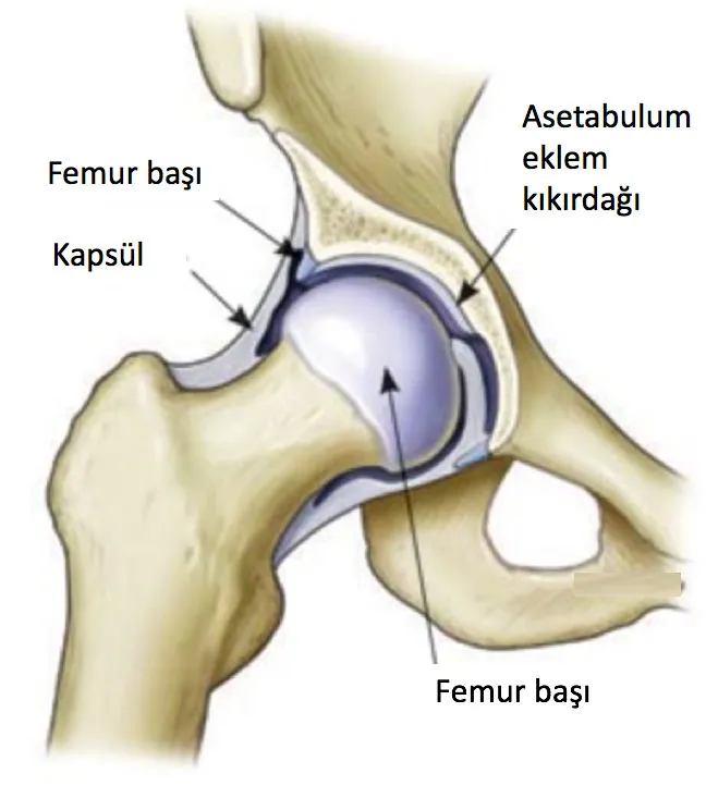
The hip joint is a ball-and-socket joint composed of the femoral head and the acetabulum. Painless movement in the joint is facilitated by the hyaline cartilage covering the joint surfaces. The hip joint capsule is a thick and strong structure that significantly contributes to the stability of the joint.
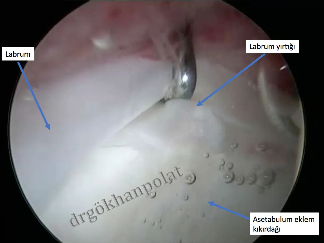
The labrum is a fibrocartilage structure that lines the rim of the acetabulum, increasing the depth of the joint and contributing to joint stability. The labrum provides a seal-like function in the joint, ensuring that synovial fluid remains in the central joint space and aiding in the survival of articular cartilage. Damage to this structure can lead to complaints such as pain, catching, and locking in the joint.
Labral tears predominantly occur in the presence of underlying bony abnormalities such as femoroacetabular impingement or acetabular dysplasia. Additionally, they can occur traumatically in athletes with hypermobility or individuals with joint laxity.
Factors causing labral tears:
Bony factors: Static overload, femoral version problems, hip deformities such as coxa valga, acetabular dysplasia, femoroacetabular impingement, dynamic impingement
Soft tissue-related: Iliopsoas impingement, laxity (collagen tissue disorders)
Traumatic: Subluxation and dislocation
Labral tears most commonly present with groin pain. Patients often describe a deep and sharp pain. The pain typically increases with activity. Activities requiring hip flexion and internal rotation, such as sitting in low positions or getting in and out of a car, tend to provoke symptoms. Rarely, patients may also experience pain in the back or lateral aspect of the hip.
The labrum tissue contains numerous free nerve endings, which is why labral tears can result in a highly painful clinical condition.
In the treatment of labral tears, aside from addressing the underlying bony problem, partial debridement of the labrum (removal of torn parts), labral repair, or labral reconstruction may be performed. The treatment decision is based on factors such as the location of the torn labral tissue, its chronicity (whether the tear is recent or old), and the tissue quality.
If the labral tissue quality is compromised (e.g., worn or calcified tissue), labral debridement may be performed. Specialized shavers and radiofrequency probes are used to remove the damaged area from the joint.
If the labral tissue quality is sufficient for healing, labral repair can be performed using bone anchor sutures placed along the acetabular rim. The bone anchors used in this repair are absorbable and dissolve within the bone after labral healing.
Figure: Appearance of the hip joint and labral tear during arthroscopy
In cases of previously failed surgical treatments or degenerative labral tears (worn and biomechanically compromised) that cannot be repaired, labral reconstruction may be performed during revision hip arthroscopy. Reconstruction can be performed using tissue harvested from another part of the body or an allograft (gracilis tendon from a cadaver).
Prof. Dr. Gökhan Polat Istanbul University Faculty of Medicine Department of Orthopedics and Traumatology
© Prof. Dr. Gökhan Polat | Orthopedics and Traumatology Specialist - All content on this website belongs to Prof. Dr. Gökhan Polat. Unauthorized copying is prohibited.