- Mon - Sat: 9:00
- Call Us: +90 (538) 894 00 91
- [email protected]

Hip Arthroscopy
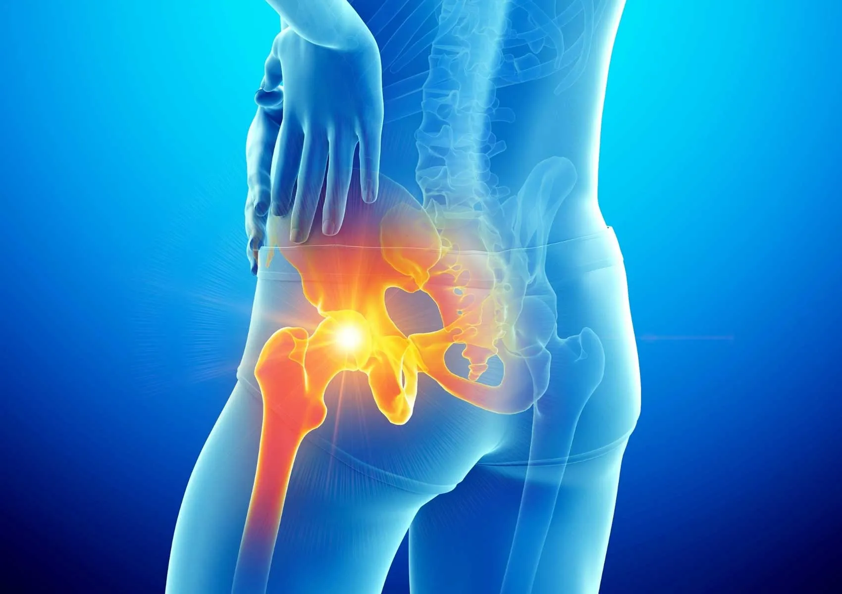
Arthroscopy is a diagnostic and treatment method used to treat joint problems by directly visualizing the structures inside the joint through small incisions.
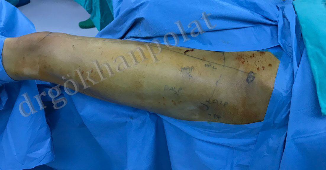
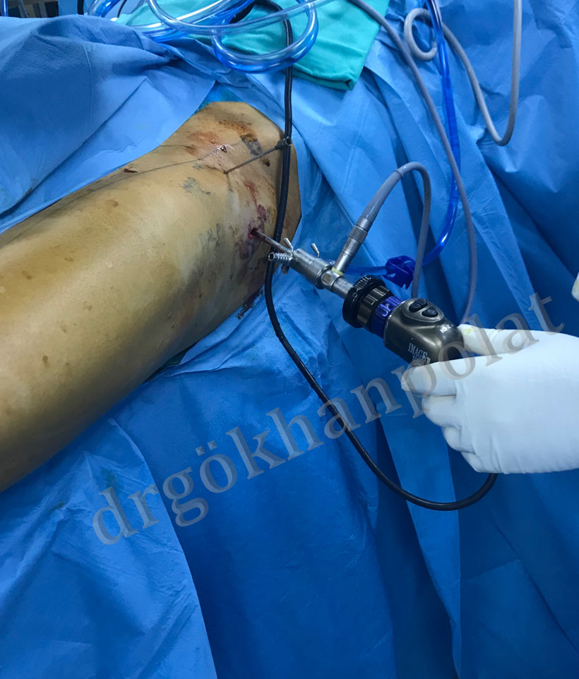
Figure 1: The joint is visualized and damaged tissues are treated through 3-4 small incisions over the hip.
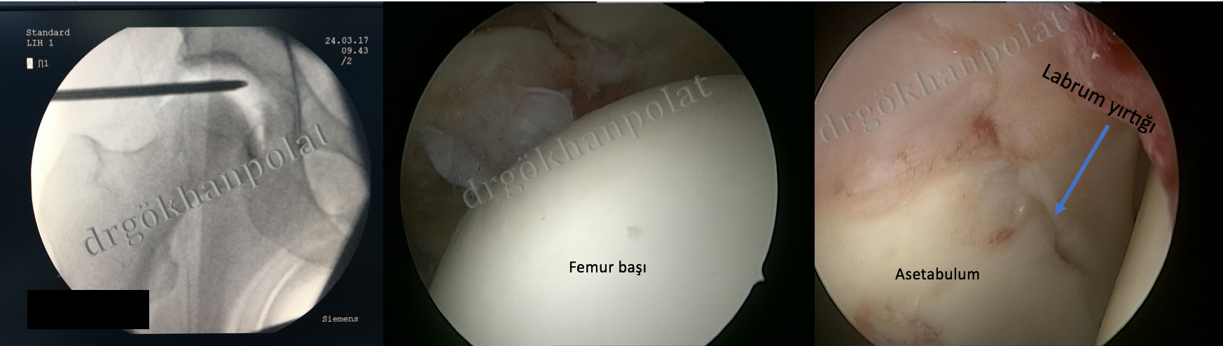
Figure 2: View of the joint interior and labrum tear during hip arthroscopy
Arthroscopic hip surgery can be used to treat many conditions inside or around the hip joint in experienced hands. Today, hip arthroscopy is most commonly used to treat conditions such as femoroacetabular impingement, labral tears, cartilage damage, iliopsoas pathologies, trochanteric bursitis, and gluteus medius tears.
Preparation for hip arthroscopy differs from standard arthroscopic surgeries due to anatomical differences in the hip joint. In addition to standard arthroscopy equipment, various devices such as traction equipment, special foot holders, and instruments are required.
In experienced hands, many conditions previously treated with open surgery can now be treated arthroscopically with similar success. Moreover, this minimally invasive surgical procedure, performed through a few small portals (entry holes), allows for faster healing and rehabilitation, enabling patients to return to their daily lives more quickly.
Hip arthroscopy is used in the treatment of many diseases.
This condition involves pathological contact and impingement between the femoral head-neck junction and the acetabulum due to morphological abnormalities in the hip joint. It can be acetabular-originated (Pincer type), femoral-originated (CAM type), or combined type (both).
Labral tears and cartilage injuries caused by impingement can lead to hip pain. This clinical condition is known as femoroacetabular impingement syndrome.
Femoroacetabular impingement is the most common condition treated with hip arthroscopy today. Through 3-4 portals, hip arthroscopy enables not only the repair of damaged structures in the joint but also the removal of the pincer lesion (acetabuloplasty) and CAM lesion (femoroplasty) that cause impingement. In experienced hands, 2- to 5-year clinical success rates are around 90%. (For more details, see “Femoroacetabular Impingement”)
The labrum is a fibrocartilaginous tissue surrounding the acetabular rim. It increases the depth of the hip joint and is crucial for joint stability and cartilage survival.
Labral tears often occur due to underlying bony abnormalities such as femoroacetabular impingement or acetabular dysplasia. They can also be seen in hypermobile athletes or individuals with ligamentous laxity.
Treatment includes addressing the underlying bony issues, partial labral debridement (removal of torn parts), labral repair, or labral reconstruction. The treatment decision is based on factors such as the location of the tear, chronicity, and tissue quality.
If the labrum has poor tissue quality (worn or calcified), debridement is performed. Special shavers and radiofrequency probes are used to remove the damaged area from the joint.
If the tissue quality is sufficient, the labrum can be repaired by applying absorbable bone anchors to the acetabular rim. These anchors dissolve inside the bone after healing.
In cases of failed previous surgeries or irreparable degenerative tears, reconstruction is performed during revision hip arthroscopy using tissue from another part of the body or allograft (gracilis tendon from a cadaver).
Cartilage damage on the acetabular or femoral head side of the hip joint can be treated arthroscopically following general cartilage repair principles. Microfracture is the most commonly used method today.
This condition presents with painful snapping during certain hip movements. A tight iliopsoas tendon can impinge on the acetabular rim or femoral head. The tendon can be released and lengthened arthroscopically at the joint level or lesser trochanter. (Iliopsoas Tenotomy)
Also known as coxa saltans, this condition causes painful snapping on the lateral side of the hip. A tight iliotibial band slides over the greater trochanter during hip movement. In patients unresponsive to physical therapy or injections, the iliotibial band can be released arthroscopically. (Iliotibial Band Release)
Commonly referred to as “Greater Trochanteric Pain Syndrome,” this clinical condition involves hip pain due to iliotibial band tightness, gluteus medius tendinosis, or bursitis. Surgical treatment may be considered for symptomatic patients who do not respond to steroid injections and rehabilitation. During surgery, the trochanteric bursa is cleaned and any associated issues are treated arthroscopically. (Trochanteric Bursectomy)
The gluteus medius muscle is responsible for hip abduction and is actively engaged during walking. Tears in this muscle or its tendon can cause hip pain and limping. In patients aged 40-60 with unresolved symptoms despite conservative treatment, tears can be repaired arthroscopically. (Arthroscopic Gluteus Medius Repair)
The synovial tissue lining the joint capsule produces synovial fluid, essential for cartilage nourishment. In certain synovial diseases (e.g., synovial chondromatosis, inflammatory arthritis), hip pain may develop. For patients whose symptoms persist despite medical treatment or due to mass effect, surgical treatment may be required. Through hip arthroscopy, the joint can be visualized, biopsy samples can be taken, and inflamed synovial tissue can be removed.
Prof. Dr. Gökhan Polat Istanbul University Faculty of Medicine Department of Orthopedics and Traumatology
© Prof. Dr. Gökhan Polat | Orthopedics and Traumatology Specialist - All content on this website belongs to Prof. Dr. Gökhan Polat. Unauthorized copying is prohibited.