"Fluid loss in the knee" is not a medical diagnosis but a popular expression, widely used in Turkey. It usually describes narrowing of the joint space on an X-ray, meaning that the cartilage lining the knee joint has thinned and worn. The real issue is not fluid "running out" but gradual wear of the cartilage that protects the joint surfaces, and treatment is aimed at that.
This article explains what so-called fluid loss really corresponds to, its symptoms, how it is diagnosed, the role of exercise and knee braces, treatment options including injections, and when to see an orthopedic specialist.
Can the knee really lose fluid?
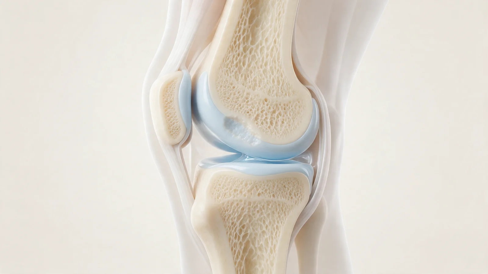
The knee joint is enclosed in a capsule containing a small amount of slippery synovial fluid, which helps the joint surfaces glide and nourishes the cartilage. In a healthy knee this amount is small, and the joint lining constantly produces and renews it. The fluid does not simply run out.
So where does the phrase come from? Cartilage is invisible on X-ray; the apparent gap between the thigh bone and shin bone is actually filled with cartilage and menisci. As cartilage thins, the gap narrows. Being told "your joint space is narrowed" was gradually understood as "the fluid in your knee has decreased". In practice, "fluid loss" usually means cartilage wear and osteoarthritis.
The opposite, fluid build-up in the knee, is a different problem caused by inflammation, injury or osteoarthritis; see our article on what fluid accumulation in the knee means.
Why does cartilage wear develop?
Joint cartilage has a limited capacity to repair itself, so accumulated load or an injury can leave lasting changes. Contributing factors include:
- Age: cartilage changes in water content and elasticity over time.
- Excess weight: increases the load on the knee.
- Previous injuries: meniscus tears, ACL injuries and fractures into the joint raise later risk.
- Alignment: bow legs or knock knees concentrate load on one compartment.
- Muscle weakness: weak thigh muscles reduce the joint's shock protection.
- Genetics and some rheumatic conditions
What are the symptoms?
- Knee pain that worsens on stairs, when squatting or after long walks
- Stiffness in the morning or after sitting for a long time
- A grinding feeling or crackling with movement
- Occasional swelling
- Difficulty fully bending or straightening the knee
- In later stages, leg deformity and changes in walking
These symptoms can also occur with other knee problems, so an examination is needed to find the cause.
How is it diagnosed?
The examination assesses where it hurts, range of motion, swelling and alignment. Standing X-rays show joint space narrowing and other osteoarthritis signs. If X-rays are still normal, or another problem such as a meniscus tear is suspected, an MRI may be requested. Reports may say "joint space narrowing", "degenerative changes" or "chondral damage"; these are the medical terms behind "fluid loss".
Treatment options
The aims are to ease pain, preserve function and help slow progression. The approach depends on the degree of wear, age, activity level and symptoms.
Exercise and weight control
Strengthening the quadriceps, hips and calves spreads the load more evenly. Straight leg raises, seated knee extensions, controlled sit-to-stands and glute bridges, together with walking, cycling and swimming, are commonly recommended. In people who are overweight, even a few kilograms of weight loss can reduce the load on the knee.
Do knee braces help?
Elastic sleeves can give some people a sense of support and short-term relief. When wear is concentrated in one compartment, an unloader brace may be used on medical advice. Wearing a brace all day for long periods can weaken the muscles, so ask your doctor which type to use and for how long. A brace does not replace exercise.
Medication and physiotherapy
Painkillers and anti-inflammatory gels can help during flare-ups but should be used under medical guidance because of stomach, kidney and heart risks. Physiotherapy ensures exercises are done correctly and progressed appropriately.
Joint injections
What patients call "putting fluid into the knee" is usually a hyaluronic acid injection. Hyaluronic acid is a natural component of joint fluid; the aim is not to top up missing fluid but to ease pain and improve movement in suitable patients, and the effect varies. PRP and corticosteroid injections may be considered in certain situations, depending on the examination and imaging.
Surgical options
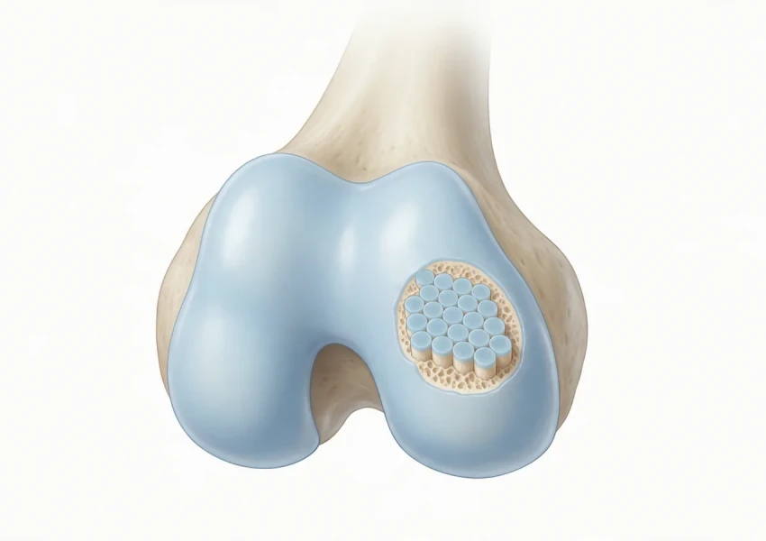
Cartilage repair techniques for limited defects in young, active people, osteotomy for malalignment and joint replacement for advanced osteoarthritis may be discussed. See our article on current cartilage damage treatments.
When should you see an orthopedic specialist?
- Knee pain lasting several weeks that limits daily activities
- Recurrent swelling
- Catching, locking or giving way
- Being unable to fully straighten or bend the knee
- Increasing leg deformity
- Pain that wakes you at night
Seek same-day care if the knee is red, hot and swollen with a fever, or if one calf becomes swollen and painful.
At Prof. Dr. Gökhan Polat's clinic, treatment for cartilage wear is planned around examination and imaging findings, the location and extent of the wear, age and activity level. Learn more on our cartilage diseases page.
In summary
- "Fluid loss in the knee" is not a medical term; it usually describes joint space narrowing, meaning cartilage wear.
- Symptoms include pain on stairs, morning stiffness, crackling and occasional swelling.
- Exercise and weight control are the foundation; braces, medication and injections are supportive.
- The right treatment is decided individually after examination and tests.
This article is for general information only; diagnosis and treatment decisions are made individually after examination and the necessary tests.
Frequently asked questions
What does fluid loss in the knee mean?
Fluid loss in the knee is a popular expression rather than a medical diagnosis. It usually describes narrowing of the joint space on X-ray, meaning the cartilage has thinned and worn, while the joint fluid itself is continuously produced by the joint lining.
How is fluid loss in the knee treated?
The foundation is exercise to strengthen the thigh and hip muscles, together with weight control. Physiotherapy, medication and joint injections such as hyaluronic acid or PRP may be added, and in advanced cases surgical options such as osteotomy or replacement are considered individually.
Should I wear a knee brace for fluid loss in the knee?
Elastic sleeves can give some people short-term relief and a sense of support, and an unloader brace may be used on medical advice when wear is concentrated in one compartment. Because wearing a brace all day for long periods can weaken the muscles, agree on how long to use it with your doctor.
Do hyaluronic acid knee injections work?
Hyaluronic acid injections can help ease pain and improve movement in suitable patients, but the effect varies from person to person and they do not rebuild worn cartilage. Whether they are appropriate is decided based on the examination and X-ray findings.
What exercises can I do for cartilage wear in the knee?
Strengthening moves such as straight leg raises, seated knee extensions, quad sets, controlled sit-to-stands and glute bridges, along with walking, cycling and swimming, are commonly recommended. It is wise to avoid deep squatting, kneeling and movements that clearly increase pain.