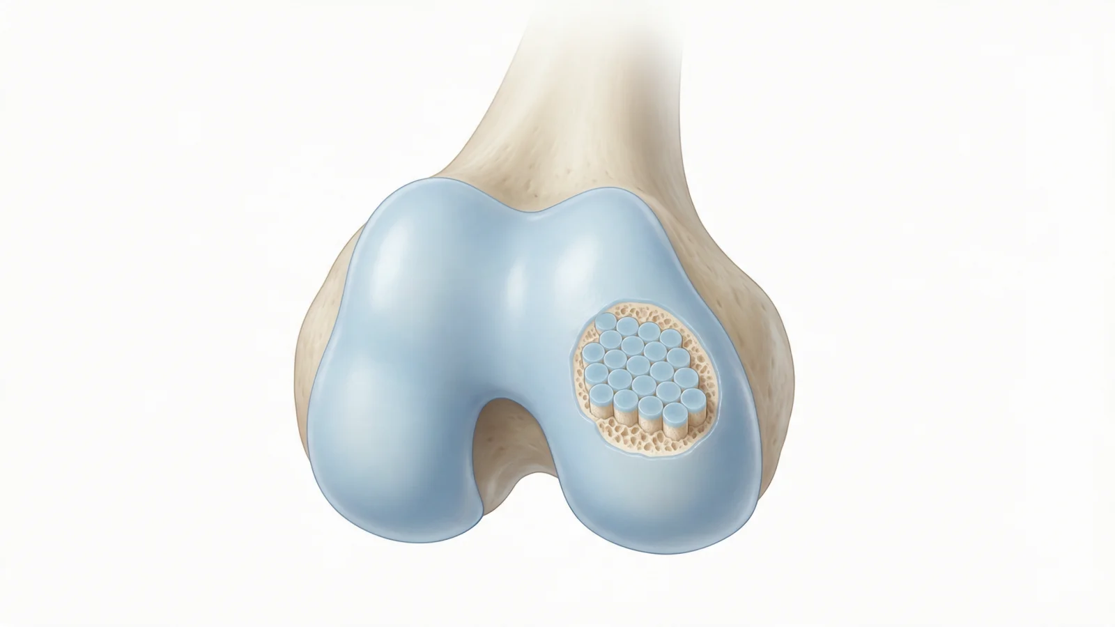
Mosaicplasty is a procedure that fills a well-defined area of damaged cartilage and underlying bone in the knee (an osteochondral defect) with small cylindrical plugs of cartilage and bone taken from a less weight-bearing part of the patient's own knee. The plugs are placed side by side like the tiles of a mosaic, hence the name. It is usually considered for young, active patients with small to medium, clearly bordered defects, and is not suitable for widespread osteoarthritis. "Cartilage transplantation" is a broader term that also includes cartilage cell implantation and techniques using donor tissue.
This article explains what a cartilage defect is, how mosaicplasty is performed, who may be a candidate, how it compares with other cartilage repair and transplant methods, the possible risks and what recovery involves.
What is a cartilage defect in the knee?
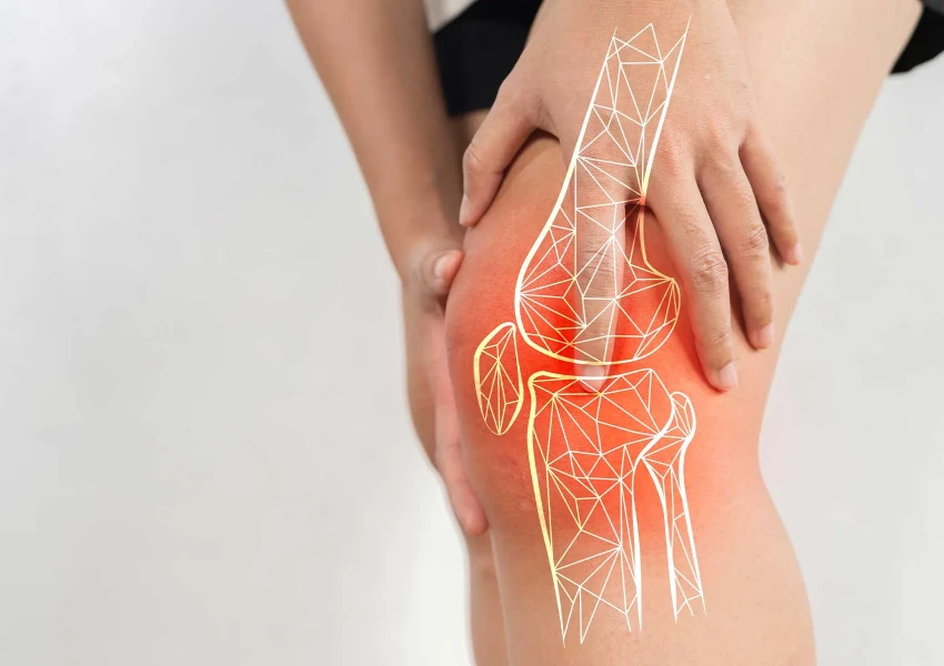
Joint cartilage is the smooth tissue covering the ends of the bones that lets the joint move with minimal friction. It has no blood supply, so its ability to heal is very limited. Sports injuries, kneecap dislocation, ligament injuries, osteochondritis dissecans (a problem with the blood supply to bone beneath the cartilage) or repeated loading can create a clearly bordered area of damage. Such focal defects are different from osteoarthritis, in which wear is spread across the joint.
A cartilage defect may cause:
- Deep knee pain with weight-bearing, stairs or sport
- Recurrent swelling after activity
- Catching, locking or a feeling that something is moving in the knee
- Noise or grinding with movement
Diagnosis is based on examination and MRI, and the size, depth and condition of the underlying bone are sometimes most clearly assessed during arthroscopy. We explain why early assessment matters in cartilage damage in the knee.
How is mosaicplasty performed?
Depending on the location and size of the defect, mosaicplasty can be done arthroscopically or through a small incision. The main steps are:
- Assessment: the joint is inspected, the defect is measured and any meniscus or ligament problems are checked.
- Preparing the recipient site: damaged cartilage and, if needed, unhealthy bone beneath it are removed by drilling neat cylindrical sockets.
- Harvesting the grafts: small cylinders of bone topped with healthy cartilage are taken from less weight-bearing areas, usually the edges of the thigh bone at the front of the knee.
- Placement: the cylinders are press-fitted into the sockets so that their surface sits flush with the surrounding cartilage. Small gaps between them gradually fill with repair tissue.
Its advantage is that the defect is filled in a single operation with the patient's own true joint cartilage, supported by bone. Its limitation is the small amount of tissue available, so it may not be enough on its own for large defects. For an overview of arthroscopic techniques, see how cartilage repair is performed with arthroscopy.
Who may be a candidate?
Suitability is decided individually based on examination, MRI and, where needed, arthroscopy. Factors that generally favour mosaicplasty include:
- A young, active patient (there is no strict age limit; the biological state of the joint matters most)
- A clearly bordered, small to medium defect in one area
- Largely healthy cartilage around and opposite the defect
- Normal leg alignment, or malalignment that can be corrected at the same time
- Stable ligaments, or ones that can be repaired in the same operation, and a preserved meniscus
Widespread osteoarthritis, cartilage loss in several areas, inflammatory joint disease and uncorrected malalignment generally make mosaicplasty unsuitable.
Other cartilage repair and transplant options
- Microfracture (marrow stimulation): small holes in the base of the defect let marrow cells form repair tissue. It is mainly used for small defects, and the tissue formed is more fibrous than true joint cartilage.
- Augmented marrow stimulation: a membrane placed over a microfracture helps hold the repair tissue in the defect.
- Autologous chondrocyte implantation (cartilage cell transplant): a small cartilage sample is taken, the cells are grown in a laboratory and implanted in a second operation. It may be considered for larger defects, but requires two stages and specialised laboratory support.
- Osteochondral allograft (donor tissue): used for large cartilage and bone defects; access to donor tissue varies between countries.
- Biological adjuncts: stem cell and PRP treatments are being studied as support for surgery in some cases. See stem cell applications in cartilage injuries.
Possible risks
- Pain or swelling where the grafts were taken (donor site symptoms)
- Incomplete healing of the plugs or a surface that is not perfectly flush
- Fluid in the joint or stiffness
- General surgical risks such as infection, bleeding and blood clots
- Symptoms returning over time and the need for further surgery
The benefit varies from person to person. The characteristics of the defect, correction of associated problems and commitment to rehabilitation all influence the outcome.
Recovery after mosaicplasty
Because the plugs take time to integrate, the post-operative period is planned carefully and tailored to the defect:
- Early weeks: weight-bearing on the operated leg is usually restricted for several weeks, with crutches, while range-of-motion exercises start early.
- Middle phase: load increases gradually and physiotherapy focuses on strengthening the thigh and hip muscles, adding stationary cycling and pool work.
- Return to sport: running and impact sports usually take several months and depend on strength, joint movement and follow-up findings.
Read about the general principles in rehabilitation after arthroscopic surgery.
When should you see an orthopedic surgeon?
- Knee pain that has persisted for weeks after a sports injury or kneecap dislocation
- Recurrent swelling after activity
- Catching, locking or a feeling of a loose fragment
- An MRI report mentioning a cartilage defect, osteochondral lesion or osteochondritis dissecans
At Prof. Dr. Gökhan Polat's clinic, the choice of treatment for cartilage defects takes into account the size and location of the defect, any meniscus, ligament or alignment problems, age and activity goals, and non-surgical options are also reviewed where appropriate. Learn more on our cartilage diseases page.
Summary
- Mosaicplasty fills a focal cartilage and bone defect in the knee with small cylindrical grafts from the patient's own knee.
- It is usually considered for young, active patients with small to medium focal defects and is not suitable for widespread osteoarthritis.
- Microfracture, cartilage cell implantation and donor tissue grafts are alternatives depending on the defect.
- Recovery takes months and outcomes vary; treatment is decided individually based on examination and imaging.
This article is for general information only; diagnosis and treatment decisions are made individually after examination and the necessary tests.
Frequently asked questions
How is mosaicplasty performed?
In mosaicplasty, small cylinders of bone topped with healthy cartilage are taken from less weight-bearing areas of the knee. They are press-fitted into sockets drilled in the damaged area so that their surface sits flush with the surrounding cartilage. Depending on the defect, it can be done arthroscopically or through a small incision.
Who is suitable for a cartilage transplant?
Cartilage transplant techniques are usually considered for young, active patients with a clearly bordered focal defect, suitable leg alignment and stable ligaments. They are generally not suitable for widespread osteoarthritis. The choice of method depends on the size and location of the defect and is made individually.
When can you walk after mosaicplasty?
After mosaicplasty, weight-bearing on the operated leg is usually restricted for several weeks and crutches are used. Load is then increased gradually based on follow-up visits. The timeline varies with the location and size of the defect.
What is the difference between mosaicplasty and microfracture?
Microfracture creates small holes in the base of the defect so that marrow cells can form repair tissue, which is more fibrous in nature. Mosaicplasty fills the defect with plugs carrying the patient's own true joint cartilage on a bone base. Microfracture is mainly used for small defects, mosaicplasty for small to medium ones.
Can cartilage damage heal on its own?
Because joint cartilage has no blood supply, its ability to repair itself is very limited, and a significant defect usually does not return to normal on its own. Small defects that cause few symptoms may, however, stay quiet for a long time with exercise and activity changes. The need for treatment is decided based on examination and MRI findings.