
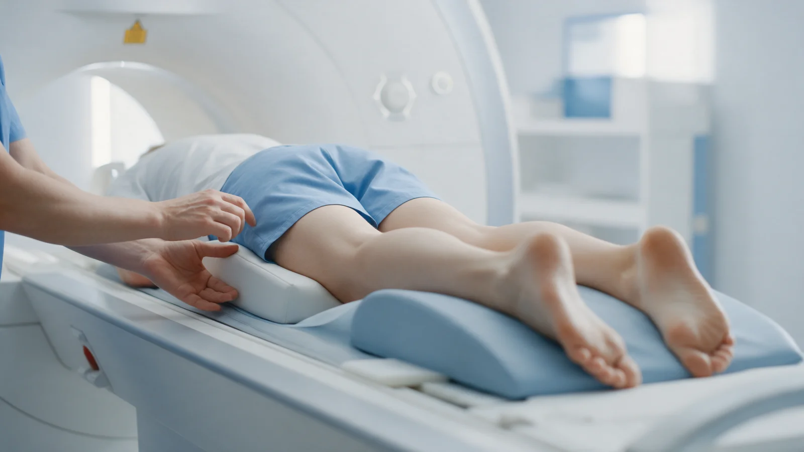
Hip MR Arthrogram: Why Contrast MRI Is Used for Labral Tears
A hip MR arthrogram is an MRI taken after contrast is injected into the joint, showing the labrum and cartilage in more detail. Learn when it is requested, how it is done and what to expect afterwards.
A hip MR arthrogram is an MRI scan performed after a small amount of diluted contrast is injected into the hip joint through a fine needle. The fluid gently distends the joint capsule and fills the space around the labrum and cartilage, outlining these structures more clearly. As a result, when a labral tear or cartilage damage is suspected, it can provide more detail than a standard hip MRI without contrast. It is not needed for every hip pain; the choice of test depends on the examination findings and the question that needs answering.
This article explains the difference between a standard hip MRI and an MR arthrogram, when a contrast study is requested, how the procedure is done step by step, how to prepare and what to expect afterwards, the possible side effects and how the results are interpreted.
How does a standard hip MRI differ from an MR arthrogram?
A standard hip MRI is taken without any injection and is very useful for many problems around the hip:
- Bone marrow oedema, stress fractures and missed fractures
- Loss of blood supply to the femoral head (avascular necrosis)
- Tendons, bursae and muscle tears on the outer hip
- Joint fluid and inflammatory changes
- Bone and soft tissue masses
The labrum, however, is thin and firmly attached to the rim of the socket. Because there is normally very little fluid inside the joint, small labral tears and early surface cartilage damage are not always clearly visible on a standard scan. In an MR arthrogram, the injected fluid seeps into a tear or between the labrum and bone, making the tear easier to see. This is why it is considered a more sensitive method for assessing labral tears and cartilage damage.
Non-contrast scans on newer high-resolution MRI machines are increasingly good at showing the labrum. In some patients a standard MRI is therefore enough, while in others an arthrogram is preferred. The choice depends on the clinical question, the equipment available and the individual patient.
When is a hip MR arthrogram requested?
- Symptoms suggesting a labral tear, such as groin pain that is worse when sitting or bending and turning the hip, catching or locking
- Signs of femoroacetabular impingement on examination and X-ray, with a need to assess the labrum and cartilage
- A standard MRI that is normal or inconclusive while symptoms still point to a problem inside the joint
- Detailed assessment of the labrum and cartilage when hip arthroscopy is being planned
- Persistent symptoms after a previous hip arthroscopy
For more on the symptoms and diagnosis of labral tears, see hip labrum tear: symptoms, diagnosis and treatment.
How is the procedure done?
- Preparation: You will be asked about allergies, medications (especially blood thinners), kidney disease, possible pregnancy and any metal implants or pacemaker. The procedure is explained and consent is obtained.
- Injection into the joint: The skin at the front of the hip is cleaned and numbed with local anaesthetic. Using fluoroscopy (live X-ray) or ultrasound guidance, a fine needle is placed in the joint and diluted contrast is injected, often together with a local anaesthetic. This step is usually short; you may feel pressure or fullness.
- MRI scan: The scan is taken shortly afterwards, while the contrast is spread through the joint. Keeping still during the scan is important for image quality, and scan time varies with the machine and protocol.
If a local anaesthetic was included, you may be asked to note how your usual hip pain changes over the next few hours. A clear reduction in pain is a valuable clue that the pain comes from inside the joint and can help guide treatment planning.
What should you watch for before and after?
Before
- Tell the team in advance if you take blood thinners, and do not stop them on your own.
- Mention any previous allergic reaction to contrast or local anaesthetics.
- Let them know if you could be pregnant.
- Report any pacemaker, metal implant or metal fragments for MRI safety.
- If possible, bring someone with you and plan not to drive yourself.
After
- You may be advised to avoid strenuous exercise, long walks and heavy lifting the following day, while continuing normal daily movement.
- A feeling of fullness or mild ache in the hip for a day or two is common and usually settles by itself.
- If redness, increasing pain or fever develops at the injection site, seek medical attention promptly.
What are the possible side effects and risks?
An MR arthrogram is generally well tolerated, but as with any joint injection there are some risks:
- Temporary pain or fullness in the hip for a day or two
- Bruising at the injection site
- Allergic reaction to the contrast or local anaesthetic (rare)
- Joint infection (very rare; sterile technique keeps the risk to a minimum)
- Temporary leg numbness from the local anaesthetic
Risks and benefits are weighed individually, and an arthrogram is requested only when the result is likely to influence treatment.
When may an MR arthrogram not be suitable?
In some situations the arthrogram is postponed, another method is chosen or extra precautions are taken:
- A skin infection or open wound around the hip or injection site
- Suspected joint infection, where the priority is to confirm the diagnosis by other means
- A previous serious allergic reaction to contrast
- Pregnancy
- A pacemaker or certain metal implants that prevent MRI
- Severe difficulty being in an enclosed space, in which case options are discussed with your doctor
Sharing this information beforehand helps the team plan the procedure safely.
How are the results interpreted?
The report may describe the labrum (whether there is a tear, its location and type), the cartilage, bone shape findings such as a cam deformity that can cause impingement, cysts next to the labrum and other associated changes. A tear in the report does not necessarily mean it is the cause of the pain or that surgery is needed, because labral tears are also seen in people without symptoms.
Results are therefore interpreted together with the examination and X-ray. For the role of imaging in impingement, see the causes and diagnosis of femoroacetabular impingement. At Prof. Dr. Gökhan Polat's clinic, imaging is always assessed alongside symptoms and examination findings, and treatment often starts without surgery and is tailored to each patient. You can read more about the labrum on our hip anatomy and labrum tears page.
When should you see an orthopedic surgeon?
- Groin pain lasting several weeks that is worse when sitting or bending the hip
- Catching, locking or sharp pain with a clicking sensation
- Ongoing hip symptoms despite a normal standard MRI
- A labral tear on your MRI report and you would like to understand your treatment options
Seek urgent care if: after an MR arthrogram you develop redness at the injection site, steadily increasing pain, fever or chills, which need same-day assessment for joint infection. Go to the emergency department for signs of an allergic reaction such as shortness of breath, a widespread itchy rash or facial swelling.
In summary
- A hip MR arthrogram is an MRI taken after contrast is injected into the joint, and it can show the labrum and cartilage in more detail.
- It is requested when a labral tear or cartilage damage is suspected and the result is likely to change treatment.
- Local anaesthetic added to the injection can give a clue as to whether the pain comes from inside the joint.
- A tear on the report is not a reason for surgery on its own; results are interpreted alongside the examination.
This article is for general information only; diagnosis and treatment decisions are made individually after examination and the necessary tests.
FAQ
-
Does a labral tear show up on MRI?
Labral tears can be seen on a standard MRI, but small tears and early cartilage damage are not always clearly visible. In an MR arthrogram, contrast injected into the joint fills the tear and can make it more obvious. Newer high-resolution machines are also increasingly good at showing the labrum without contrast.
-
Is a hip MR arthrogram painful?
The skin is numbed with local anaesthetic before the injection, and most people feel pressure or fullness rather than sharp pain. A mild ache or fullness in the hip for a day or two afterwards is common and usually settles on its own. How uncomfortable it feels varies from person to person.
-
What is the difference between a hip MRI and an MR arthrogram?
A standard hip MRI is taken without any injection and is valuable for many conditions such as bone marrow oedema, stress fractures, avascular necrosis and tendon or bursa problems. In an MR arthrogram, contrast is first injected into the joint, allowing the labrum and cartilage to be assessed in more detail.
-
What should I do after an MR arthrogram?
You may be advised to avoid strenuous exercise, long walks and heavy lifting the next day, while carrying on with normal daily movement. If possible, do not drive yourself on the day. Seek medical attention promptly if you notice redness, steadily increasing pain or fever at the injection site.
-
If my MRI shows a labral tear, do I need surgery?
Not necessarily. Labral tears are also found in people without symptoms, and most symptomatic tears are treated without surgery first. The decision about surgery is based not on the MRI image but on symptom severity and duration, examination findings and the overall condition of the joint.