
When Do You Need a Knee MRI? X-Ray, Ultrasound and MRI Compared
When is a knee MRI needed and when might it be unnecessary? What X-ray, ultrasound and MRI can and cannot show in the knee, whether a meniscus tear shows on X-ray and how to prepare for the scan.
A knee MRI shows the menisci, ligaments, cartilage and bone marrow in detail, but it is not needed for every case of knee pain. It is usually requested when the examination suggests a meniscus tear or ligament injury, when the knee locks or gives way, or when pain persists without a clear cause after several weeks of appropriate treatment. X-rays are better for bones and arthritis, and ultrasound for superficial tendons and fluid; the menisci and cruciate ligaments do not show up on X-ray.
This article explains what X-ray, ultrasound and MRI can and cannot show in knee pain, when a knee MRI is genuinely useful, when it may be unnecessary, how to prepare for the scan and how a healthy meniscus differs from a torn one on MRI.
Why imaging is not the first step
The first and most important step in assessing knee pain is the examination. How the pain started, where it is felt, which movements make it worse and the results of specific clinical tests often narrow down the likely cause considerably. Imaging is then used to confirm that impression, rule out other causes or refine the treatment plan.
An MRI done too early or without a clear question can sometimes cause confusion. Meniscal wear and small tears are common even in middle-aged and older people with no symptoms, and such findings may be unrelated to the pain yet lead to unnecessary worry or procedures. The right question is not "should I have an MRI?" but "will the MRI change my treatment?"
What do X-ray, ultrasound and MRI show?
X-ray
- Shows: bone structure, fractures, signs of arthritis (joint space narrowing, bone spurs), leg alignment and kneecap position.
- Does not show: menisci, ligaments, the cartilage itself or tendons.
- Note: standing X-rays are more informative for arthritis because they show the joint space under load.
Can a meniscus tear be seen on X-ray? No. Even so, an X-ray is often requested when a meniscus tear is suspected, because it shows whether arthritis, a loose bone fragment or another bone problem is present, which can change the treatment plan.
Ultrasound
- Shows: tendons close to the surface (patellar and quadriceps tendons), fluid in and around the joint, Baker's cysts and bursitis.
- Limits: cannot adequately show deep structures such as the cruciate ligaments, the inner meniscus or cartilage, and results depend heavily on the operator.
- Advantages: no radiation, quick, allows assessment during movement and can guide injections.
MRI
- Shows: menisci, anterior and posterior cruciate ligaments, collateral ligaments, cartilage, bone marrow edema, hidden and stress fractures, impaired blood supply to bone, cysts and masses.
- Limits: relatively expensive and time-consuming, difficult for people with claustrophobia, and some metal implants and devices may prevent scanning.
- Note: no radiation is involved.
For a broader comparison, see our article on imaging methods used in orthopedic diseases.
When is a knee MRI needed?
- Rapid swelling after a twisting sports injury with a suspected ACL tear
- Mechanical symptoms such as locking, catching or being unable to straighten the knee
- Repeated episodes of the knee giving way
- Pain without a clear cause after examination and X-ray that does not respond to several weeks of appropriate treatment
- Assessing cartilage or ligament damage after a kneecap dislocation
- Suspected stress fracture, bone marrow edema or impaired blood supply to bone
- Planning surgery, to define the type and location of a tear and any associated damage
For more on meniscus tear symptoms, read what is a meniscus tear and what are the symptoms?
When might a knee MRI be unnecessary?
- Advanced arthritis already visible on X-ray: MRI usually adds detail about known wear without changing the treatment decision.
- New, mild pain without injury: it often settles with a few weeks of relative rest and exercise, so this is tried first.
- Superficial tendon or bursa problems: ultrasound often gives enough information.
These are general principles; if the examination points elsewhere, your doctor may request an MRI earlier.
What does a healthy meniscus look like on MRI?
A healthy meniscus appears as a smooth, uniformly dark triangle, or a bow-tie shape on side views. Reports often say something like "the menisci show normal signal and morphology". Bright areas within the meniscus are called increased signal: if they do not reach the surface they usually indicate wear (degeneration), while signal reaching the joint surface is interpreted as a tear.
When is contrast or MR arthrography used?
Most knee MRIs are done without contrast and give enough information. In some situations, contrast is injected into a vein or directly into the joint, for example to tell whether a previously repaired or operated meniscus has torn again, to see certain cartilage problems more clearly or to assess cysts and masses. Your doctor decides which technique is needed based on the examination.
How to prepare for an MRI
- Tell the staff in advance about a pacemaker, cochlear implant, drug pump, metal fragments or shrapnel. Most orthopedic implants are MRI-compatible, but you should still mention them.
- Mention claustrophobia beforehand; open MRI or other ways to help can be discussed.
- Tell the team if you might be pregnant.
- Knee MRI usually does not need contrast; if it does, you will be asked about kidney function and allergies.
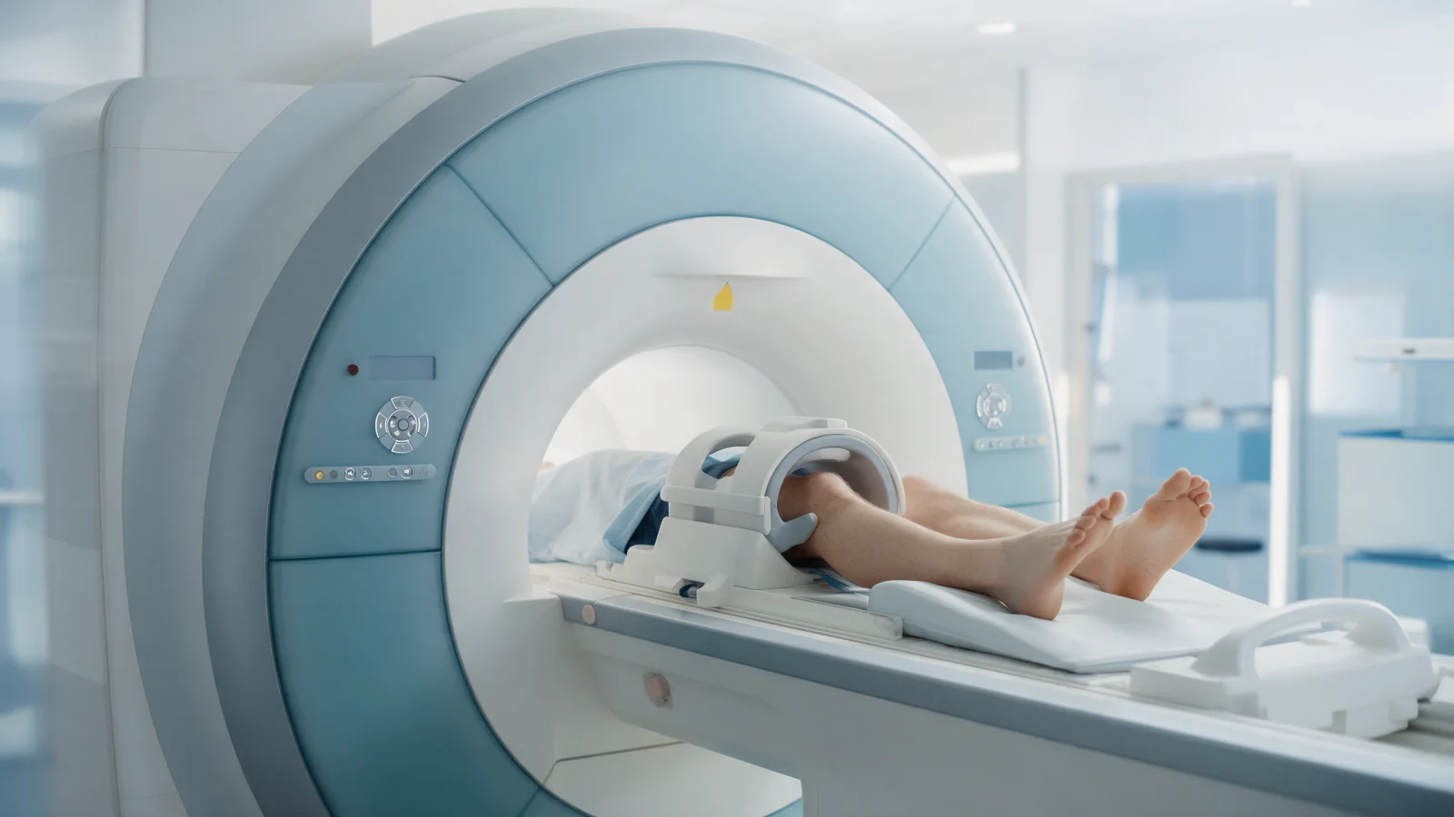
- Keeping your leg still during the scan matters for image quality. The scan usually takes around half an hour or less.
- Bring previous MRIs and X-rays for comparison.
How does the MRI affect treatment?
An MRI is only one part of the treatment decision. The same finding can lead to different plans in two people with different symptoms and goals. A repairable traumatic meniscus tear in a young athlete may need arthroscopic repair, while a degenerative tear without mechanical symptoms in a middle-aged person is often managed with physiotherapy. If arthroscopy is planned, the MRI also helps the surgeon prepare. To learn when arthroscopic techniques are used, see our arthroscopic surgery page.
When should you see an orthopedic surgeon?
- Swelling soon after an injury, or a "popping" sensation
- Locking, catching or inability to straighten the knee
- The knee giving way when walking
- Inability to bear weight
- Night pain or pain that does not ease with rest
- Pain that does not improve, or keeps worsening, after a few weeks of self-care
Urgent: redness, warmth and swelling with fever, or obvious deformity after a fall, need same-day emergency assessment.
At Prof. Dr. Gökhan Polat's clinic, the need for imaging is decided based on the examination, and MRI results are interpreted from the images themselves alongside your symptoms. Treatment plans are individual, and response to treatment varies from person to person.
Summary
- X-ray shows bone and arthritis, ultrasound shows superficial tendons and fluid, and MRI shows menisci, ligaments, cartilage and bone marrow.
- Menisci and cruciate ligaments are invisible on X-ray, but X-rays still provide important information.
- Knee MRI is most useful for suspected ligament or meniscus injury, mechanical symptoms and unexplained persistent pain.
- MRI findings do not decide treatment on their own; they are interpreted alongside the examination.
This article is for general information only; diagnosis and treatment decisions are made individually after examination and the necessary tests.
FAQ
-
Does a meniscus tear show up on X-ray?
No. The meniscus is made of cartilage and does not show on X-ray; MRI is the main test for meniscus tears. An X-ray is still often requested because it can show arthritis, loose bone fragments or other bone problems that affect the treatment plan.
-
How long does a knee MRI take?
A knee MRI usually takes around half an hour or less, depending on the scanner and the sequences needed. Keeping the leg still during the scan is important for image quality.
-
Should I have an X-ray or an MRI first for knee pain?
In most cases the examination comes first, followed by an X-ray if needed. MRI is usually chosen when a meniscus or ligament injury is suspected, when there are mechanical symptoms, or when pain persists without a clear cause after several weeks of treatment.
-
What does a healthy meniscus look like on MRI?
A healthy meniscus appears as a smooth, uniformly dark triangle, or a bow-tie shape on side views. The report usually states that the menisci show normal signal and morphology.
-
Can I have an MRI if I have metal in my knee?
Most orthopedic implants are MRI-compatible, although image quality may be reduced near the metal. Devices such as pacemakers, cochlear implants or drug pumps may prevent scanning. Always tell the team before the scan.