
Hip Arthroscopy: Procedure, Risks, and Recovery Guide
Hip joint conditions can significantly affect daily quality of life and cause limitations in movement. With advances in technology, minimally invasive methods have become prominent in treating these problems. Hip arthroscopy is a sophisticated surgical technique performed through small incisions to visualize, diagnose, and treat intra-articular hip issues. Compared to traditional open surgery, it involves smaller cuts which can positively influence the recovery process.
This guide provides detailed information about what hip arthroscopy is, indications for its use, the surgical procedure, and recovery. Our goal is to offer a scientific and reliable resource for patients exploring treatment options, helping them make an informed decision. This method is often applied in cases of sports injuries and structural hip disorders and can achieve successful outcomes with proper patient selection and rehabilitation.
What is Hip Arthroscopy?
Hip arthroscopy involves using a thin, illuminated instrument equipped with a camera called an "arthroscope" to inspect the inside of the hip joint. This technique allows the surgeon to examine the joint structures closely and treat damaged tissues with minimal invasiveness. Instead of large incisions around the joint, only small entry points of a few millimeters are used.
Hip Joint and Labrum Anatomy
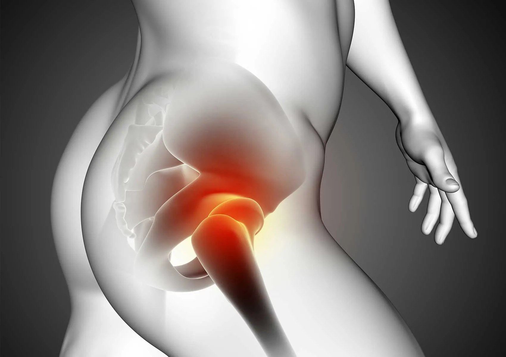
The hip joint is a ball-and-socket articulation where the head of the femur (thighbone) fits into the socket (acetabulum) of the pelvis. A critical component for joint stability is the "labrum," a ring of cartilage that deepens the socket to hold the femoral head more securely. It acts as a seal preserving joint fluid and cushioning the joint surfaces. The complex anatomy of the hip joint and the location of the labrum are illustrated in the diagram below.
Anatomical abnormalities or tears in this area can cause pain and movement restrictions. For detailed information, please visit our hip anatomy and labral tear page. Additionally, our general hip joint disorders guide covers other factors affecting joint health.
Definition and Indications of Hip Arthroscopy
Hip arthroscopy serves both diagnostic and therapeutic purposes. It is used when radiological imaging is insufficient to definitively diagnose intra-articular pathologies but is now primarily applied as a treatment method. Procedures such as cartilage repair, removal of loose bodies, alleviation of impingement syndromes, and cleansing of inflamed joint lining can be conducted with this minimally invasive approach. The reduced trauma to surrounding tissues leads to less postoperative pain and enables earlier initiation of rehabilitation.
When is Hip Arthroscopy Recommended?
Hip arthroscopy is not suitable for all causes of hip pain. It is typically chosen for cases that do not respond to conservative treatments (medication, physical therapy, rest) and where mechanical problems are identified. An orthopedic specialist evaluates the patient’s history, physical exam, and imaging before recommending this surgery.
Femoroacetabular Impingement
One of the most frequent reasons for hip arthroscopy is femoroacetabular impingement (FAI) syndrome. This condition arises due to structural bony overgrowths on the hip joint bones leading to abnormal contact and pinching during movement. Over time, this can cause labral tears and cartilage damage. In arthroscopic surgery for FAI, bone excesses are reshaped to restore the joint's range of motion and relieve impingement. More about femoroacetabular impingement syndrome can be found on our site.
Labral Tears and Other Conditions
Labral tears may result from sports injuries or underlying anatomical issues and can cause sharp groin pain. Arthroscopy allows for labral repair (suturing) or debridement if the tissue cannot be repaired. Additional indications for hip arthroscopy include synovial chondromatosis (loose cartilage fragments), ligamentum teres injuries, peritrochanteric tendon problems (snapping hip), and early-stage osteoarthritis.
The Hip Arthroscopy Surgical Procedure
The surgical process includes all stages from preoperative preparation to discharge. Careful planning of each step is necessary for a successful outcome. Procedures under the hip arthroscopy service follow international standards.
Preoperative Preparation
Before surgery, a thorough assessment of the patient’s overall health is performed along with necessary blood tests and an anesthesia consultation. Medications, especially blood thinners, are adjusted under medical supervision. Key pre- and postoperative guidelines are summarized in the checklist below.
Surgical Steps
Hip arthroscopy is typically performed under general or spinal anesthesia. The patient is positioned on a specialized traction table, where gentle pulling is applied to the leg to create space within the hip joint. This ensures safe instrument insertion. The procedure is carried out under fluoroscopic (X-ray) guidance. The key stages are illustrated in the following infographic.
The surgeon inserts the arthroscope and surgical tools through small incisions to thoroughly inspect the joint, identify damaged areas, and execute the planned intervention (repair, trimming, cleaning). Surgery duration ranges from 1 to 3 hours depending on the complexity.
Early Postoperative Period
After surgery, patients are usually monitored overnight in the hospital. Pain is managed with medications, and antibiotics may be administered to prevent infections. To reduce the risk of blood clots, compression stockings and blood-thinning injections may be recommended. Patients are generally mobilized with crutches on the same or following day.
Potential Risks of Hip Arthroscopy
Like all surgeries, hip arthroscopy carries potential risks, although they are uncommon. Possible complications include infection, bleeding, injury to blood vessels or nerves, and temporary nerve numbness due to traction (pudendal nerve compression), which usually resolves spontaneously. Additionally, insufficient resection of bone impingement or postoperative adhesions may necessitate revision surgery. With experienced surgical teams, these risks are minimized.
Recovery Process and Rehabilitation
Recovery is a critical factor influencing surgical success. The duration and course depend on patient-specific factors such as age, physical condition, and the type of procedure (labral repair versus debridement). An overview of the recovery timeline is shown below.
Physical Therapy and Exercise Program
Passive exercises begin from the first day post-surgery. Rehabilitation protocols aim to maintain joint mobility and enhance muscle strength. In some cases, platelet-rich plasma (PRP) therapy may be used as an adjunct to promote tissue healing. Supervised exercises focus on strengthening the muscles around the hip to reduce joint load. Illustrated guides of recommended exercises are provided below.
Returning to Daily Activities
Most patients use crutches for 2 to 4 weeks, although this period may be extended if cartilage repair was performed. Return to desk-based work is often possible within 2 to 3 weeks, whereas physically demanding jobs may require 3 to 4 months. Gradual reintroduction to sports, especially those involving running and jumping, requires full restoration of muscle strength and balance. Similar to recovery protocols following anterior cruciate ligament surgery, patience and discipline are essential.
When performed with proper indication and expertise, hip arthroscopy can enable patients to resume an active and pain-free lifestyle. Consult your doctor to discuss all aspects and develop a tailored treatment plan.
This content is intended for informational purposes only and does not constitute professional medical advice, diagnosis, or treatment. For any health-related questions, please consult a qualified healthcare professional or medical facility. The information provided on this site is not a substitute for examination or testing by a physician. While the content is based on current scientific data, medical knowledge is constantly evolving; therefore, you should consult your doctor for the most accurate and up-to-date information. Do not delay seeking medical care or disregard professional advice based on this content.
FAQ
-
What is hip arthroscopy and when is it recommended?
Hip arthroscopy is a minimally invasive surgical technique involving small incisions to diagnose and treat joint issues. It is commonly used for conditions like labral tears, cartilage damage, or impingement syndrome. A specialist will determine the most appropriate treatment for your specific condition.
-
What are the risks associated with hip arthroscopy surgery?
As with any surgery, hip arthroscopy carries potential risks such as infection, bleeding, nerve injury, blood clots, or reactions to anesthesia. Although these risks are uncommon, it is important to discuss them thoroughly with your doctor before the procedure.
-
How long is the recovery period after surgery and what should I be mindful of?
Recovery time varies depending on your overall health and the procedure performed, typically ranging from a few weeks to several months. Consistent participation in physical therapy, following your doctor's instructions, and avoiding excessive strain are essential for a successful recovery.
-
Is hip arthroscopy a painful surgery?
You will not feel pain during the surgery as it is performed under general anesthesia. Postoperative discomfort or mild pain is normal and can be managed with pain medication. Your doctor can help you develop an appropriate pain management plan.
-
When can I resume normal activities after hip arthroscopy?
The timeline for returning to normal activities depends on the extent of the surgery and your recovery progress. Most patients gradually return to daily activities and sports under the guidance of their doctor and physical therapist. Full recovery often takes between three to six months; consult your doctor for a personalized plan.