
Instruments and Techniques Used in Arthroscopy
Arthroscopy is a minimally invasive surgical method used for diagnosing and treating joint diseases. The specialized instruments used and the techniques applied determine the success of the operation. This article examines the basic tools used in arthroscopy and the most common surgical techniques in detail.
Arthroscopy is one of the widely used minimally invasive surgical methods in orthopedics and traumatology for diagnosing and treating joint diseases. This method is performed by inserting a small camera and various slender surgical instruments into the joint. Due to the small incisions required for arthroscopic procedures, patients experience less pain, a faster recovery period, and a reduced risk of complications.
The instruments used in arthroscopy are highly specialized to ensure the procedure is conducted effectively and safely. Additionally, the applied techniques are carefully adapted according to the specific joint and pathology. This article will comprehensively review the most commonly used instruments in arthroscopic surgery and the fundamental techniques employed.
Arthroscopic Instruments
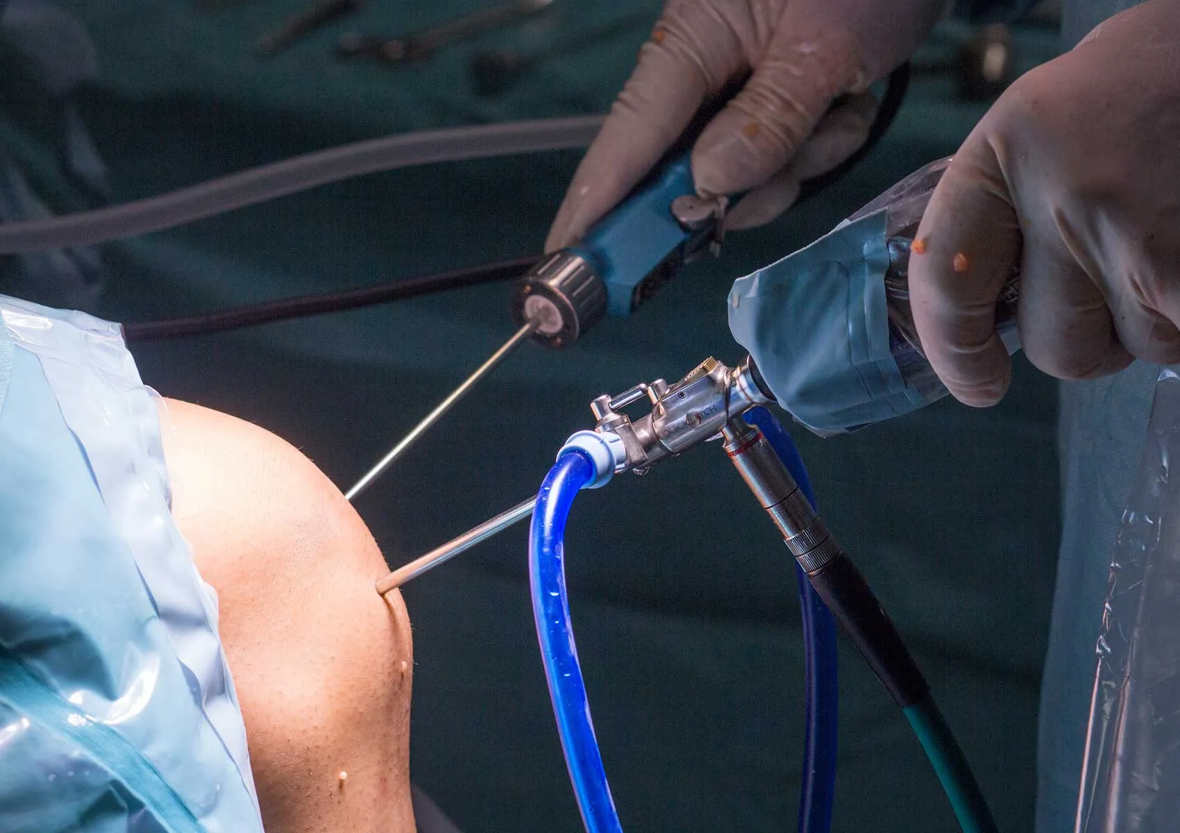
Arthroscope:
The arthroscope is generally an optical device with a diameter of 4 millimeters and a 30-degree viewing angle. It is inserted into the joint through a small incision, allowing illumination and visualization of the interior of the joint. Modern arthroscopes provide high-resolution images, with some models offering different viewing angles, such as 70 degrees.
Camera System and Monitor:
The arthroscope is attached to a camera system, and images are displayed in real-time on a monitor. This system guides the surgeon during the operation and ensures a clear view of the surgical field.
Instrument Ports (Trocars):
Besides the portal through which the arthroscope is inserted, various thin tools used for tasks such as suturing, cutting, and scraping are introduced into the joint. These ports allow different instruments to be utilized based on the procedure.
Fluid Pump and Injection System:
During arthroscopic surgeries, the joint cavity is inflated and cleaned with fluid. The fluid pump clarifies the view inside the joint, minimizes bleeding, and expands the surgical field.
Surgical Instruments:
- Probe: Used to examine intra-articular tissues.
- Shaver (cleaning device): Used to remove damaged tissues and clean bone surfaces.
- Blades and Scissors: Used to cut torn tissues.
- Electro-cautery: Generates electrical current for bleeding control.
- Suture Passer and Needles: Facilitate passing sutures during labrum or meniscus repairs.
- Powered Instruments: Preferred for shaping and scraping bone or cartilage.
Arthroscopic Techniques
Diagnostic Arthroscopy:
This is the initial step performed for the definitive diagnosis of joint problems. The internal joint structures are examined with the camera, and damages are detected. If necessary, treatment follows this phase.
Meniscus Repair and Partial Meniscectomy:
Meniscus tears are common in the knee joint. Depending on the location and shape of the tear, either repair or partial removal of the meniscus is performed. These techniques are chosen based on the severity of the damage.
Anterior Cruciate Ligament Reconstruction:
In cases of anterior cruciate ligament injury, the ligament is reconstructed using a tendon graft. This is done arthroscopically through minimal incisions, resulting in a faster healing process.
Hip Arthroscopy:
Used for diagnosing and treating hip conditions such as femoroacetabular impingement, labrum tears, and gluteus medius tears. These techniques reduce tissue damage compared to open surgery.
Cartilage Repairs:
In cases of cartilage damage, techniques such as microfracture, autologous chondrocyte implantation, and tissue engineering can be applied arthroscopically. These methods contribute to the regeneration of the joint surface.
Joint Debridement and Synovectomy:
Removal of damaged tissues and inflamed synovial tissue helps reduce pain and inflammation. This is especially preferred in athletes following chronic injuries.
Emerging Techniques and Robot-Assisted Arthroscopy:
In recent years, navigation systems and robot-assisted devices have enhanced the accuracy of arthroscopic surgeries and reduced complications.
Advantages of arthroscopy include a low risk of infection due to its minimally invasive nature, less bleeding, early mobilization, and a shorter hospital stay. However, the surgeon's experience, the quality of instruments used, and technique choices directly affect the success of the outcomes. Arthroscopy holds an increasingly important role, particularly in the treatment of sports injuries and joint diseases within the fields of orthopedics and traumatology.
FAQ
-
Which joints are arthroscopy most commonly performed on?
The knee, shoulder, and hip joints are the most frequently treated with arthroscopy. Additionally, arthroscopic surgery can also be performed on joints such as the elbow, wrist, and ankle.
-
What is a shaver used in arthroscopic surgery, and what does it do?
A shaver is a small, electrically powered motorized device that cuts and removes damaged or worn tissues during arthroscopy. It cleans the surgical field and accelerates tissue healing.
-
What is the recovery process after arthroscopic surgeries?
Because arthroscopic surgery is minimally invasive, the recovery process is typically fast. Patients can return to their daily activities in a short time, although physical therapy may be required depending on the joint and type of procedure performed.
-
How is arthroscopy applied in the treatment of femoroacetabular impingement?
In femoroacetabular impingement, arthroscopy is used to correct the narrowed bone areas, repair or remove the labrum. These procedures improve hip function and reduce pain.
-
How are arthroscopic instruments sterilized?
All arthroscopic instruments are sterilized before surgery according to specific sterilization protocols using autoclaves or chemical disinfectants. This process is vital to minimize the risk of infection.