
What Is Acetabular Dysplasia? Symptoms and Treatment Options
A Silent Structural Threat to the Hip Joint
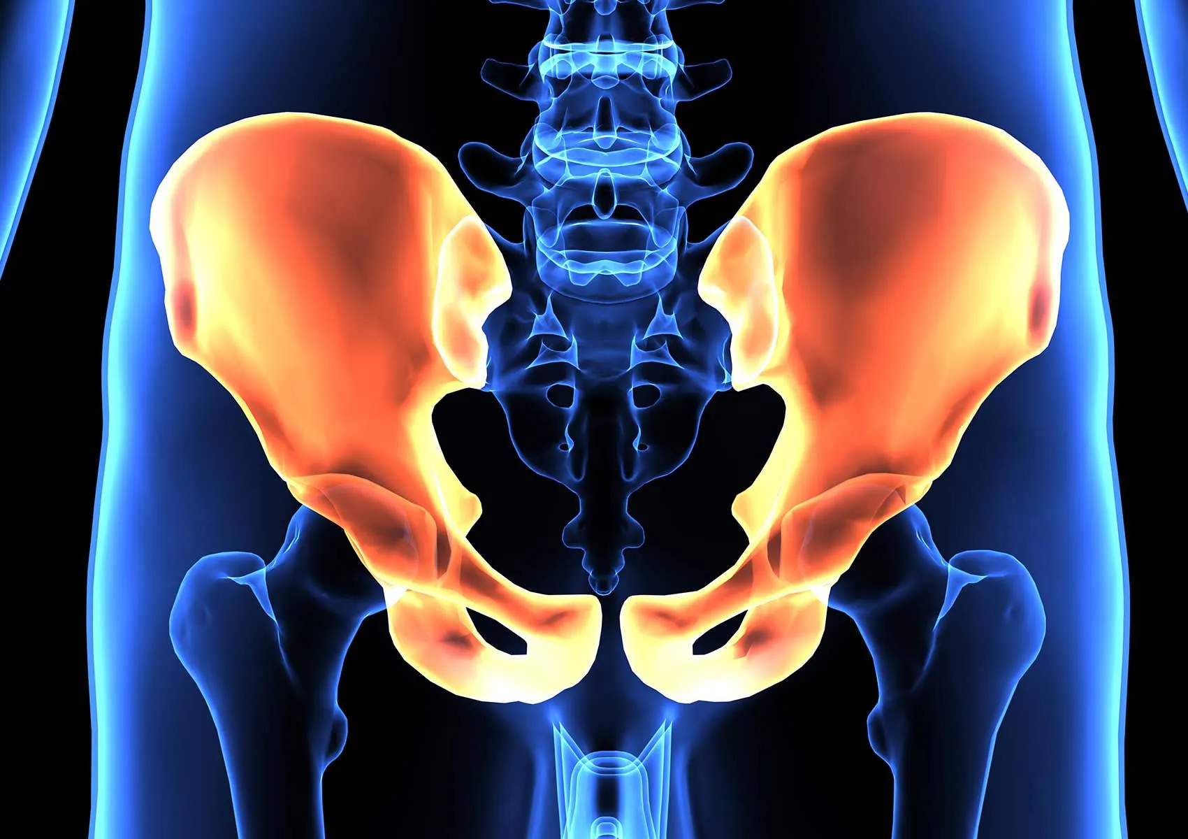
Acetabular dysplasia is a condition in which the acetabulum (hip socket) is underdeveloped or shallow, resulting in inadequate coverage and support of the femoral head. This structural imbalance impairs the stability of the hip joint and can lead to labral tears, cartilage damage, and early-onset hip osteoarthritis if left untreated. While some cases are detected in infancy, mild forms may go unnoticed until adolescence or early adulthood, often presenting as unexplained groin or hip pain.
In this article, we will explain what acetabular dysplasia is, how it presents, how it is diagnosed, and which treatment options—from physical therapy to surgery—can help preserve joint health and function.
Why Is the Acetabulum-Femur Relationship Important?
A healthy hip joint functions like a ball-and-socket system: the femoral head (ball) fits snugly into the acetabulum (socket), providing smooth movement and even distribution of forces. In acetabular dysplasia, this relationship is altered. The socket is too shallow or misaligned, which leads to increased stress on the labrum and cartilage.
Over time, this mechanical overload may cause the labrum to tear and the cartilage surface to deteriorate. This process gradually reduces joint function and increases pain, potentially resulting in osteoarthritis at a relatively young age.
What Causes Acetabular Dysplasia?
The condition is typically congenital, meaning it is present at birth due to improper development of the hip joint during fetal growth. Many cases are detected early through newborn screening and hip ultrasound. However, mild or borderline dysplasia may not be identified until adulthood.
Risk factors include:
- Female sex (more common in girls)
- Family history of hip dysplasia
- First-born babies with difficult or breech deliveries
- Swaddling or tight wrapping during infancy
- Limited hip mobility in early childhood
Some individuals with subclinical dysplasia may remain symptom-free until they become more physically active or start experiencing wear-and-tear on the joint.
What Are the Symptoms?
Symptoms of acetabular dysplasia depend on the severity of the condition and may evolve gradually over time. Common signs include:
- Groin or lateral hip pain, often activity-related
- Discomfort when standing or walking for extended periods
- Sharp or catching pain when rising from a seated position
- Clicking or popping sensations in the hip
- Pain when climbing stairs or running
- Limping or a feeling of leg length discrepancy in advanced cases
These symptoms can become more pronounced as joint damage progresses, and they may be mistaken for other conditions like a sports injury or muscle strain.
How Is It Diagnosed?
Diagnosis begins with a detailed clinical examination, assessing hip range of motion, gait, and pain location. Standard X-rays are essential to evaluate the acetabular shape, femoral head coverage, and load distribution.
Additional imaging may include:
- MRI: Helps detect labral tears and cartilage damage
- MR arthrography: Involves injecting contrast into the joint for detailed labrum assessment
- CT scan: Provides 3D visualization for preoperative planning or complex cases
Accurate diagnosis is crucial for determining the appropriate treatment strategy.
What Are the Treatment Options?
The treatment plan depends on factors such as the degree of dysplasia, patient age, activity level, and presence of structural damage. Treatment falls into two broad categories: non-surgical and surgical.
1. Conservative (Non-Surgical) Management:
- Physical therapy to strengthen surrounding hip muscles
- Activity modification (avoiding high-impact sports)
- Anti-inflammatory medications for pain relief
- Guided exercise programs to enhance hip stability
- Lifestyle adjustments to reduce joint overload
These approaches are more suitable for mild dysplasia without labral damage or for individuals not yet ready for surgery.
2. Surgical Intervention:
For patients with persistent symptoms or moderate to severe dysplasia, especially in younger individuals, surgery may be necessary.
The most effective surgical method is:
Periacetabular Osteotomy (PAO):
This procedure involves cutting and repositioning the pelvic bone to rotate the acetabulum and increase coverage of the femoral head. It restores load balance, protects the labrum and cartilage, and significantly delays the onset of osteoarthritis.
PAO is particularly effective for young, active patients and offers the possibility of preserving the natural hip joint for many years.
What Happens If It's Left Untreated?
Untreated acetabular dysplasia can lead to progressive degeneration of the hip joint. The imbalance in load distribution increases stress on the labrum and cartilage, resulting in labral tears and eventual joint surface breakdown.
This deterioration leads to early-onset hip osteoarthritis, which may require total hip replacement at a much younger age than typical. Early diagnosis and appropriate treatment can prevent or significantly delay this outcome.
FAQ
-
Is acetabular dysplasia always present at birth?
Yes, it is typically congenital, though mild cases may go undiagnosed until adulthood.
-
Does every case require surgery?
No. Mild or asymptomatic cases may be managed conservatively. Surgery is reserved for symptomatic or progressing cases.
-
Can acetabular dysplasia worsen over time?
Yes. If untreated, it can lead to labral tears and early osteoarthritis due to uneven joint loading.
-
How long is the recovery after PAO surgery?
Initial recovery takes 3–6 months, but full return to activity may take up to 9–12 months depending on rehabilitation.
-
Can I return to sports after treatment?
Yes. With proper treatment and rehabilitation, low-impact sports can usually be resumed. High-impact sports should be evaluated case by case.