
Hip Arthroscopy: Comprehensive Guide and FAI Syndrome Treatment
Hip arthroscopy has gained significant importance in orthopedics in recent years thanks to technological advancements. It plays an effective role in diagnosing and treating hip pain, particularly in young and active individuals. Performed with smaller incisions compared to traditional open surgery, this technique aims to accelerate patients’ return to daily life. It is widely preferred for managing various hip pathologies, especially femoroacetabular impingement (FAI) syndrome, as part of joint-preserving surgery.
Introduction to Hip Arthroscopy
The hip joint is one of the deepest and most complex joints in the body. Surgical treatment of problems in this region previously required large incisions, but today arthroscopic methods offer a more comfortable experience.
What is hip arthroscopy?
Hip arthroscopy is a minimally invasive surgical procedure used to visualize, diagnose, and treat internal structures of the hip joint. Also known as “keyhole surgery,” this method involves inserting a pencil-thin camera (arthroscope) and surgical instruments through small incisions into the joint. This allows the surgeon to examine joint cartilage, the labrum, and surrounding soft tissues in detail. For further information on indications and procedures, visit hip arthroscopy indications.
When is it indicated?
This procedure is typically considered for hip pain unresponsive to conservative (non-surgical) treatments. The most common indications include:
- Femoroacetabular impingement (FAI) syndrome
- Labral tears
- Removal of loose bodies inside the joint
- Synovial diseases
- Cartilage damage
- Peri-hip tendon issues (e.g., snapping hip)
The Hip Joint and FAI Syndrome
Anatomical abnormalities of the hip joint can lead to mechanical problems and pain over time, with FAI syndrome being a primary cause.
Hip joint anatomy
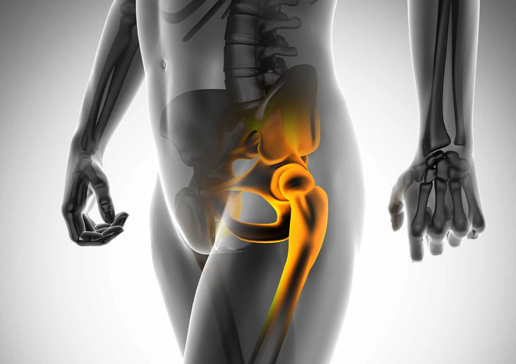
The hip is a ball-and-socket joint formed between the head of the femur (thigh bone) and a socket in the pelvis called the acetabulum. The joint surfaces are covered by smooth cartilage, and the labrum, a fibrocartilaginous structure, surrounds the socket providing stability. Understanding healthy hip anatomy is essential for grasping the nature of related disorders. For more on this, see the importance of hip joint anatomy.
The diagram below visualizes the normal hip anatomy and how FAI syndrome affects the joint.
What is femoroacetabular impingement (FAI)?
FAI occurs when abnormal shapes of the hip bones (femur and acetabulum) cause them to rub against each other during movement. This abnormal contact can damage the labrum and cartilage over time. FAI is classified into three types: Cam (bump on the femoral head), Pincer (acetabular overcoverage), and Mixed. For detailed information, visit femoroacetabular impingement explained.
The comparison table below summarizes the different FAI types and their features.
Symptoms and Diagnosis of FAI Syndrome
Early diagnosis is critical to prevent further joint damage. Patients usually report pain that worsens with specific movements.
Symptoms
The hallmark symptom of FAI syndrome is groin pain, which may radiate to the outer thigh or buttocks. Pain often increases with prolonged sitting, driving, squatting, or sports. Mechanical symptoms like catching, locking, or clicking of the hip can also be present. Since symptoms overlap with other hip conditions, differential diagnosis is important. See hip joint disease and treatments for related conditions.
Diagnostic methods
Diagnosis starts with a thorough physical examination. The physician will perform specific maneuvers, such as impingement tests, to reproduce pain and identify the source. Imaging is essential to confirm the diagnosis and plan surgery, including:
- X-rays: Reveal bony abnormalities (Cam or Pincer lesions).
- Magnetic Resonance Imaging (MRI): Shows labral tears and cartilage damage in detail.
The image below demonstrates pre- and post-operative MRI scans for hip arthroscopy cases.
Hip Arthroscopy Procedure
When surgical treatment is decided, understanding the procedure is a key concern for patients.
Surgical steps
Hip arthroscopy is performed on a traction table, where slight pulling force is applied to the leg to widen the joint space and allow instrument access. Under fluoroscopic guidance (live X-ray), the surgeon inserts instruments and the arthroscope into the joint. The labral tears are repaired, excess bone is shaved, and cartilage surfaces are smoothed. For complete procedural details, please visit hip arthroscopy surgery details.
The flowchart below summarizes the surgical steps.
Duration and anesthesia
The operation is generally performed under general or spinal anesthesia. Surgery duration varies depending on the interventions but typically lasts between 1 to 2 hours. Most patients are discharged on the same day or the following day.
Postoperative Recovery and Rehabilitation
A well-structured rehabilitation program is vital for a successful surgical outcome.
Physical therapy and exercise programs
Use of crutches and movement restrictions are common in the first days after surgery. Physical therapy begins early to help restore range of motion and strengthen muscles around the hip. Rehabilitation after arthroscopic treatment of femoroacetabular impingement should be tailored individually. For more details, see arthroscopic treatment rehabilitation.
The table below outlines the stages of recovery and therapy programs.
Recovery timeframe and precautions
Full recovery and return to sports vary by patient. Desk jobs can usually be resumed within 2-3 weeks, while high-intensity sports may require 4-6 months. It is critical to follow the surgeon’s instructions closely and avoid sudden or strenuous movements during recovery.
Risks and Alternatives to Hip Arthroscopy
As with any surgery, hip arthroscopy carries potential risks and may not be suitable for every patient.
Possible complications
Although generally safe, rare complications include temporary nerve damage from traction (numbness), infection, bleeding, or joint stiffness. Patients with advanced osteoarthritis may not benefit as expected from arthroscopy. Therefore, joint-preserving surgery options and risks should be discussed thoroughly with the patient.
Alternative treatments
For patients unsuitable or unwilling to undergo surgery, alternatives include activity modifications, anti-inflammatory medications, physical therapy, and intra-articular injections. For information on biological therapies, see PRP treatment for hip joint.
In conclusion, hip arthroscopy is an effective method to relieve hip pain and preserve joint function when performed on appropriately selected patients by experienced surgeons.
This content is for informational purposes only and does not substitute professional medical advice, diagnosis, or treatment. Always consult your physician or a qualified healthcare provider with any health-related questions. Do not delay or disregard obtaining medical advice based on information from this site. Joint-preserving surgery patient selection and treatment planning must be individualized by a healthcare professional.
FAQ
-
What is hip arthroscopy and when is it used?
Hip arthroscopy is a minimally invasive surgical procedure used to diagnose and treat problems within the hip joint. It is commonly performed for conditions such as femoroacetabular impingement (FAI), labral tears, and cartilage damage. Consult a specialist for detailed information and a personalized assessment.
-
What are the symptoms of femoroacetabular impingement (FAI) syndrome?
Symptoms of FAI include groin pain, limited hip movement, a catching sensation, and discomfort that worsens after prolonged sitting. These symptoms often intensify with activity. It is important to see an orthopedic specialist for an accurate diagnosis.
-
How is hip arthroscopy performed and how long does it take?
Hip arthroscopy is performed using small incisions through which specialized instruments and a camera are inserted. The surgeon repairs or corrects the affected areas. The procedure duration varies depending on complexity but typically lasts 1 to 2 hours.
-
What is the recovery process after surgery and what should be considered?
Recovery after surgery varies based on the patient's condition and the specific procedure performed. It usually involves physical therapy and exercise programs. Full recovery can take several months, and following your surgeon's instructions is essential.
-
What are the risks and success rates of hip arthroscopy?
Hip arthroscopy is generally a safe procedure but carries rare risks such as infection, nerve damage, or joint stiffness. Success rates are high, with most patients experiencing pain relief and improved function. Discuss your personal risks and expectations with your doctor.