
Avascular Necrosis of the Hip: Early Signs and Treatment
Avascular necrosis develops when blood supply to the femoral head is disrupted. Learn the risk factors, early symptoms, MRI diagnosis, staging and the joint-preserving options considered before collapse.
Avascular necrosis (AVN) is the death of part of a bone because its blood supply has been disrupted, and in the hip it most often affects the head of the thigh bone (the femoral head). It is also called osteonecrosis. In its early stages it may cause only mild groin pain and can look normal on X-ray, so it is easily missed. Yet a diagnosis made before the femoral head collapses matters, because that is when joint-preserving treatments can still be considered.
This article explains why avascular necrosis develops, who is at higher risk, the early symptoms of femoral head AVN, the role of MRI, how the disease is staged and the treatment options, from joint-preserving procedures to hip replacement.
What is avascular necrosis, and why is the hip so often affected?
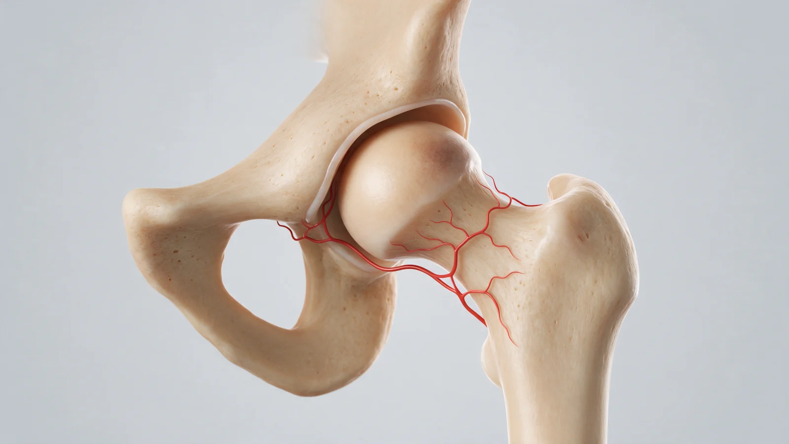
Bone is living tissue that depends on a constant blood supply. The vessels that feed the femoral head are small and few, and most of them run along the femoral neck. If they are damaged or blocked, or blood flow drops for another reason, part of the femoral head stops receiving enough blood and the bone cells in that area die.
Over time the dead bone loses its ability to carry load. If the disease progresses, the bone beneath the cartilage collapses, the normally round femoral head loses its shape and secondary arthritis develops in the joint. AVN is sometimes loosely called "bone melting" in everyday language, but it is a different condition from osteoporosis, which thins bone throughout the skeleton.
Who is at risk?
Avascular necrosis most often affects adults between about 30 and 50, and in a significant proportion of patients both hips are involved. In some people no cause is found, but the following increase the risk:
- Corticosteroid use: High-dose or long-term steroids are among the most widely recognised risk factors. This includes steroids used for rheumatic diseases, asthma, organ transplants, some blood disorders and severe infections.
- Heavy, long-term alcohol use
- Hip injuries: Femoral neck fractures and hip dislocations can directly damage the vessels of the femoral head.
- Certain blood and metabolic conditions: Sickle cell disease, clotting disorders, Gaucher disease and connective tissue diseases such as lupus.
- Other factors: Radiotherapy, some chemotherapy drugs, decompression sickness in divers and smoking.
Not everyone who takes steroids develops AVN, but new groin pain in someone who has taken them should prompt a doctor to keep this possibility in mind. Never stop steroid treatment on your own; that decision should be made with the doctor who prescribed it.
What are the symptoms of femoral head AVN?
In the earliest stage there may be no symptoms at all. When symptoms appear, they typically include:
- A deep ache in the groin, sometimes felt in the buttock or front of the thigh
- Pain that increases with weight bearing, walking and climbing stairs
- Later, pain at rest and at night
- Reduced hip movement, especially internal rotation, with difficulty putting on socks or sitting cross-legged
- A limp
In some people the pain is referred to the knee. If the femoral head collapses, pain can increase noticeably. Because these symptoms overlap with hip osteoarthritis, impingement and labral tears, only examination and imaging can tell them apart.
How is it diagnosed, and why is MRI so important?
The examination assesses hip range of motion, which movements reproduce the pain and the way you walk, and asking about risk factors is an important part of the assessment. Two main imaging methods are used:
- X-ray: Usually the first test, but it can be completely normal in early disease. In more advanced stages it may show density changes, a fracture line under the cartilage (the "crescent sign") and collapse.
- MRI: The most sensitive method for detecting early AVN. It can show the disease when the X-ray is still normal and reveals the size and location of the affected area. Because AVN is often bilateral, the other hip is usually assessed on the same MRI.
A CT scan may occasionally be used to check for subtle collapse. The diagnosis is confirmed by combining the examination with the imaging findings.
Why does the stage determine treatment?
AVN is staged according to imaging findings. Several detailed classification systems exist, but the key question for a patient is simple: has the femoral head started to collapse?
- Pre-collapse: There is an area of dead bone, but the femoral head is still round. This is when joint-preserving options are considered.
- Collapse: The bone beneath the cartilage has begun to give way and the joint surface is no longer smooth.
- Advanced: Arthritis has developed as a result of the collapse and the socket is also affected.
The size of the affected area and whether it lies in the weight-bearing part of the femoral head also influence how the disease behaves. Small lesions away from the weight-bearing zone may progress slowly, while larger ones carry a higher risk of collapse.
Joint-preserving treatment and other options
Treatment is tailored to the stage, the size of the lesion, age, activity level and the underlying cause. In general, the options include:
- Managing risk factors: Stopping alcohol and smoking where possible, and reviewing steroid treatment with the prescribing doctor.
- Non-surgical care and offloading: Small lesions without symptoms may be monitored. During painful periods, crutches to reduce load, pain relief and physiotherapy may be used. Some medications are being studied for slowing progression, but the evidence is limited.
- Core decompression: In the pre-collapse stage, small channels are drilled into the affected area to reduce pressure inside the bone and encourage new blood vessels. Some centres combine it with cell-based products from the patient's own bone marrow or with bone grafts. For background on these approaches, see our cellular therapies (PRP, stem cell) page.
- Osteotomy and vascularised bone grafts: More extensive operations used in selected younger patients to shift load onto healthy bone or support the affected area.
- Total hip replacement: Once the femoral head has collapsed and arthritis has developed, hip replacement is usually considered to relieve pain and restore movement.
The aim of joint preservation is to keep your own hip for as long as possible and delay the need for replacement, but results depend on the stage and size of the lesion and are not the same for everyone. Our article on whether joint-preserving surgery can postpone replacement covers the wider picture. At Prof. Dr. Gökhan Polat's clinic, treatment is planned around the MRI findings, stage, age and personal goals. You can read about other conditions affecting the hip on our hip joint diseases page.
When should you see an orthopedic surgeon?
- New groin or hip pain in someone who has taken, or is taking, steroids
- Groin pain that increases with weight bearing in someone with a history of heavy alcohol use
- Groin pain months or years after a hip fracture or dislocation
- Hip pain lasting several weeks, present at night or causing a limp
- New pain in the other hip in someone already diagnosed with AVN on one side
Seek urgent care if: the pain suddenly becomes much worse and you cannot bear weight, as this may mean collapse or fracture of the femoral head. Hip pain with fever and chills, especially in someone taking immune-suppressing medication, needs same-day assessment.
In summary
- Avascular necrosis is the death of bone in the femoral head caused by loss of blood supply; it is not the same as osteoporosis.
- Steroid use, heavy alcohol intake and hip injuries are the main risk factors, and both hips are often affected.
- X-rays may be normal early on; MRI is the most sensitive test for early diagnosis.
- Diagnosis before collapse allows joint-preserving options to be considered; advanced disease usually leads to hip replacement.
This article is for general information only; diagnosis and treatment decisions are made individually after examination and the necessary tests.
FAQ
-
What causes avascular necrosis?
Avascular necrosis occurs when the vessels supplying the femoral head are damaged or blood flow drops, causing bone tissue to die. High-dose or long-term steroids, heavy alcohol use, hip fractures and dislocations, sickle cell disease and clotting disorders are the main risk factors. In some people no clear cause is found.
-
What are the symptoms of avascular necrosis of the femoral head?
The most common symptom is a deep groin ache that increases with weight bearing and walking. As the disease progresses, pain may be felt at rest and at night, internal rotation becomes limited and a limp may develop. There may be no symptoms early on, so new groin pain in someone with risk factors should be assessed.
-
Does avascular necrosis show up on X-ray?
In the early stages the X-ray can be completely normal. In more advanced disease, density changes and collapse of the femoral head become visible. MRI is the most sensitive test for early diagnosis and also shows the size of the affected area and the condition of the other hip.
-
Can avascular necrosis be treated without surgery?
Small lesions without symptoms may be monitored with risk factor control and reduced loading during painful periods. However, non-surgical measures have limited ability to stop progression. Before collapse, joint-preserving surgery such as core decompression may be considered, while hip replacement is discussed once collapse and arthritis have developed.
-
Can avascular necrosis affect both hips?
Yes, avascular necrosis affects both hips in a significant proportion of patients, and sometimes the other side has no symptoms yet. For this reason, the other hip is usually assessed with MRI at diagnosis and follow-up is planned accordingly.