
Kneecap Dislocation: First Aid, Recurrence and MPFL Surgery
Why does the kneecap dislocate? What to do straight away, the factors that raise the risk of recurrence, stabilisation exercises, and when MPFL reconstruction surgery is recommended.
A kneecap dislocation (patellar dislocation) happens when the kneecap slides completely out of its groove at the front of the thighbone, almost always towards the outside of the knee. It usually causes sudden pain, a visibly misshapen knee and rapid swelling, typically during a twisting movement. Sometimes the kneecap pops back by itself; sometimes it has to be put back in an emergency department. Most first-time dislocations are treated without surgery, but repeated dislocations, or a piece of cartilage breaking off inside the joint, may lead to surgery such as MPFL reconstruction.
This article explains why the kneecap dislocates, what to do straight away, what raises the risk of it happening again, non-surgical treatment and exercises, and what MPFL surgery involves and when it is recommended.
Why does the kneecap dislocate?
The kneecap glides up and down in a groove at the front of the thighbone as the knee bends. Its stability depends on the shape of the bones and on the surrounding ligaments and muscles. The most important soft-tissue restraint on the inner side is the medial patellofemoral ligament (MPFL), which is stretched or torn in most dislocations.
Dislocation usually happens when the body twists over a planted foot, the knee collapses inwards, or the kneecap takes a blow from the side. It is more common in dance, football, basketball, volleyball and gymnastics, and in people with an anatomical predisposition it can occur with fairly simple movements.
Who is most at risk?
Kneecap dislocation is most common in teenagers and young adult athletes. Factors that can increase the risk of a first or repeat dislocation include:
- A shallow groove (trochlear dysplasia) for the kneecap to sit in
- A high-riding kneecap (patella alta)
- An outward attachment of the kneecap tendon, which increases the pull towards the outside
- Generalised joint laxity
- Knock knees and weak hip muscles
- A previous dislocation
Young people with several of these features are more likely to dislocate again. We cover other knee problems in young athletes in our article on orthopedic risks in athletic children.
First aid: what to do if your kneecap dislocates
- Do not try to force the kneecap back: untrained attempts can damage cartilage and bone. It may slip back as the knee is slowly straightened, but do not force it.
- Support the knee in the most comfortable position with a pillow or folded clothing.
- Apply cold: a towel-wrapped ice pack can help limit swelling and pain.
- Go to an emergency department, even if the kneecap has gone back into place by itself.
In hospital, the kneecap is put back with pain relief if needed and an X-ray is taken. The rapid swelling after a dislocation is usually due to bleeding inside the joint; our article on fluid in the knee explains the possible causes of a swollen knee.
Diagnosis: why an MRI is often needed
The examination assesses how mobile the kneecap is, whether pushing it outwards causes apprehension, tenderness along the MPFL and the overall stability of the knee. X-rays show fractures and the position of the kneecap.
MRI is particularly important because, as the kneecap dislocates and relocates, a fragment of cartilage or bone can break off the back of the kneecap or the thighbone. Loose fragments can cause catching and locking and may need surgery. MRI also shows where the MPFL is torn, the shape of the groove and any other injuries.
Non-surgical treatment and exercises after a first dislocation
If there is no loose fragment, first-time dislocations are usually treated without surgery, with:
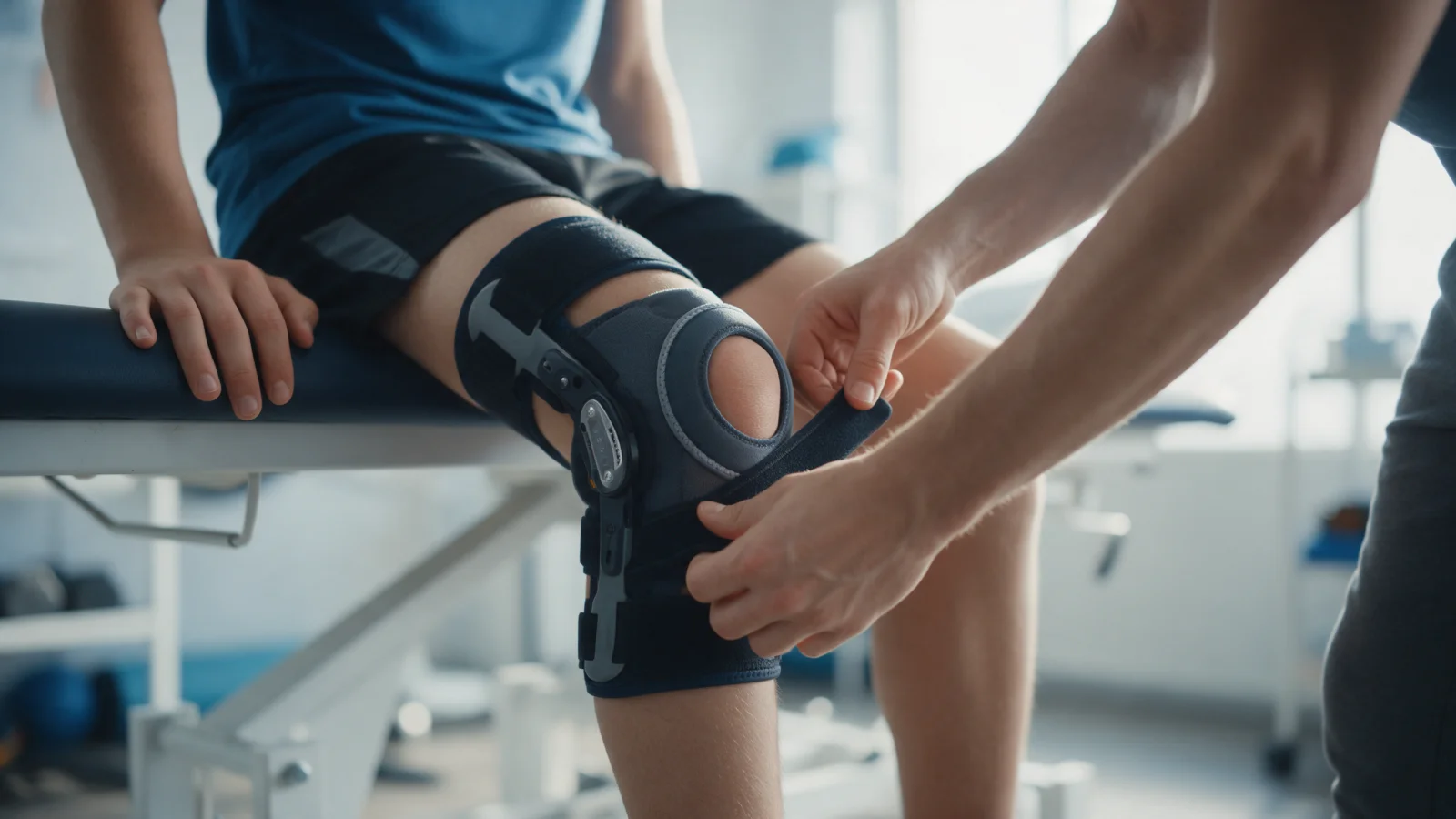
- A protective knee brace at first and crutches for a short time if needed
- Control of swelling and pain
- Gradual range-of-motion work within the limits your doctor sets
- A rehabilitation programme to strengthen the thigh, hip and core muscles
Exercises commonly included in kneecap stabilisation programmes are:
- Quadriceps sets: tightening the thigh muscle with the knee straight for a few seconds
- Straight leg raises with the knee locked
- Side-lying leg raises and clamshells to strengthen the hip muscles that stop the knee collapsing inwards
- Partial squats within a comfortable range, keeping the knees from drifting inwards
- Balance training such as standing on one leg
It is wise to plan the timing and dose of exercise with a physiotherapist after examination. Our article on strengthening the muscles around the knee explains why this matters. Return to sport varies from person to person and is usually planned once strength, balance and confidence in the knee have been regained.
What is MPFL surgery and who needs it?
MPFL reconstruction rebuilds the ligament that holds the kneecap on the inner side, using a graft taken from one of your own tendons. The aim is to restore the restraint that stops the kneecap sliding outwards. It is usually done through small incisions and is often combined with arthroscopic assessment of the joint.
MPFL surgery may be considered for:
- Repeated dislocations despite appropriate rehabilitation
- Ongoing instability that limits daily life or sport
- A cartilage or bone fragment that needs to be fixed or removed, in which case the ligament can be addressed at the same time
When there are marked anatomical risk factors, additional procedures such as an osteotomy to move the bony attachment of the kneecap tendon may be combined with MPFL reconstruction. In children with open growth plates, the technique is adapted accordingly. After surgery a brace and crutches are used for a period, followed by staged physiotherapy; return to sport usually takes several months and varies between individuals. For more on ligament surgery, see our article on arthroscopic ligament repairs.
When should you see an orthopedic surgeon?
- Any time the kneecap has come out of place, even if it went back by itself
- Rapid swelling and being unable to bear weight
- Catching, locking or a feeling that something is moving inside the knee
- Repeated episodes of the kneecap feeling as if it is about to slip
- Pain or swelling that has not settled weeks after a dislocation
At Prof. Dr. Gökhan Polat's clinic, kneecap instability is assessed by considering examination and imaging findings, the number of dislocations, anatomical risk factors, age and activity level together. For an overview of knee conditions, see our knee joint diseases page.
In summary
- A kneecap dislocation usually happens when the kneecap slips outwards during a twist, and the MPFL is often torn.
- Do not force the kneecap back; even if it relocates by itself, it needs to be assessed.
- Most first dislocations are treated without surgery using a brace and a strengthening programme.
- Repeated dislocations or a loose fragment in the joint may call for surgery such as MPFL reconstruction.
This article is for general information only; diagnosis and treatment decisions are made individually after examination and the necessary tests.
FAQ
-
What should you do if your kneecap dislocates?
Do not try to force the kneecap back. Support the knee in the most comfortable position, apply cold and go to an emergency department. Even if the kneecap has relocated by itself, an examination and X-ray are needed because a fragment of cartilage or bone may have broken off.
-
Will a dislocated kneecap happen again?
It can in some people. Young age, a shallow groove, a high-riding kneecap and general joint laxity can all increase the likelihood. A strengthening programme may help reduce the risk, and surgery is considered for repeated dislocations.
-
What is MPFL surgery?
MPFL reconstruction rebuilds the ligament that holds the kneecap on the inner side, which is often torn during a dislocation, using a graft from one of your own tendons. It is usually recommended for repeated dislocations or ongoing instability, and may be combined with bony realignment procedures when needed.
-
What exercises help an unstable kneecap?
Quadriceps sets, straight leg raises, side-lying leg raises, clamshells, partial squats and balance training are commonly used. They aim to strengthen the thigh and hip muscles that stabilise the kneecap. The programme should be tailored to the individual after examination.
-
When can you return to sport after MPFL surgery?
A brace and crutches are used for a while after surgery, followed by staged physiotherapy. Return to sport usually takes several months and is planned around strength, range of motion and confidence in the knee. The timeline depends on the procedures performed and the individual.