Imaging and Follow-up After Osteotomy
Effective monitoring of the recovery process in patients after osteotomy requires regular imaging and follow-ups. This article discusses the preferred imaging methods post-osteotomy, radiological evaluation, clinical follow-up, and details the monitoring involved during rehabilitation.
Osteotomy is a controlled surgical cut made in the bone to correct a desired deformity and is widely used in the treatment of many orthopedic conditions. Its aim is to correct malalignment in the hip, knee, and other joints or to prevent the progression of cartilage damage. Regular imaging and follow-ups are essential for ensuring a successful postoperative course and preventing complications.
The purpose and application areas of osteotomy influence both surgical planning and the follow-up process. In particular, conditions such as femoroacetabular impingement, acetabular dysplasia, or correction of malalignment in the knee require close evaluation of bone healing, implant positioning, and surrounding structures after surgery.
Imaging Methods After Osteotomy
After osteotomy, the imaging techniques commonly used can be broadly divided into three groups: standard radiography (X-ray), computed tomography (CT), and magnetic resonance imaging (MRI).
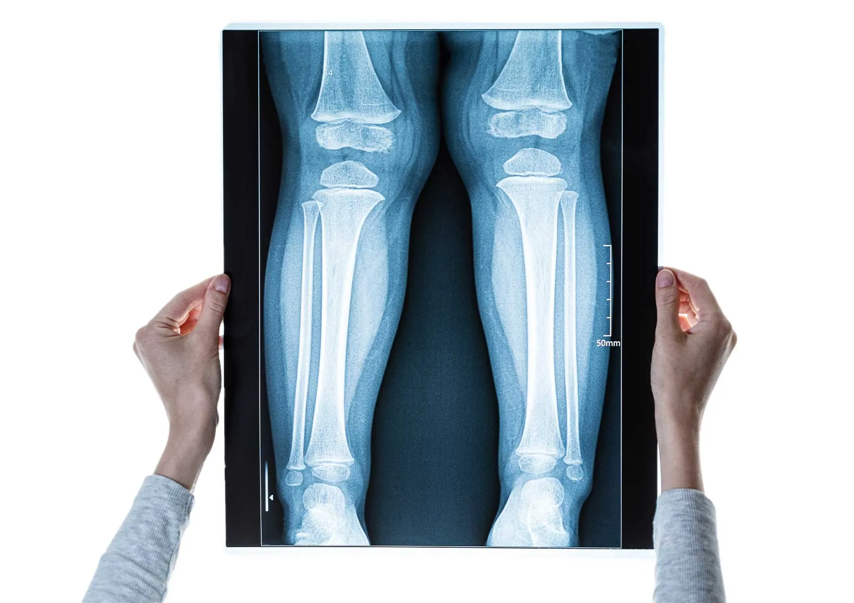
Radiography is the first and routine imaging method post-osteotomy. It is used in the early postoperative period to assess the positioning of implants, monitor the osteotomy line, and track the level of bone healing. Especially anteroposterior (AP) and lateral views serve as the fundamental reference images. X-rays are often taken at 6 weeks, 3 months, 6 months, and 1 year following surgery.
Computed tomography is preferred to reveal detailed structural changes in the bone. It has significant advantages in correcting complex deformities and thoroughly assessing bone healing. Additionally, it clearly demonstrates how well the implant fits into the bone and whether any displacement has occurred. CT scans are generally used beyond routine follow-ups, especially when investigating pathology related to patient complaints.
Magnetic resonance imaging excels in soft tissue evaluation and is useful for detecting problems in the ligaments, cartilage, or muscle tissues around the osteotomy site. It is particularly preferred for differential diagnosis in cases of infection, muscle tears, or nerve complications at the osteotomy region.
Radiological Evaluation and Follow-up Process
In the early postoperative period, evaluation focuses on implant positioning and the stability of the osteotomy line. Early calcification signs in bone healing usually appear on X-rays within the first six weeks. Complete bone healing occurs between 3 to 6 months. During this time, careful monitoring is necessary for implant loosening or recurrence of any deformity.
During follow-up visits, radiological comparisons are made to observe bone density and union progress along the osteotomy line. For patients at risk of delayed union or nonunion, additional imaging may be required. CT scans can be requested for further diagnosis in such cases.
Physical Examination and Clinical Follow-ups
Clinical examination is as important as imaging after osteotomy. At every examination, the patient's level of pain, joint range of motion, weight-bearing capacity, and functional status must be evaluated. Early mobilization without delay is targeted following osteotomy surgeries; therefore, physical examinations are critical for tracking the patient’s progress in recovery.
Clinical signs such as swelling, infection indicators, or restricted movement are noted during follow-ups. These examinations are typically scheduled at the first week, sixth week, third month, sixth month post-surgery, and at later dates if necessary.
Monitoring and Management of Complications
Potential complications after osteotomy must be identified and managed promptly. Common issues include infection, implant loosening, failure of bone union (nonunion), and nerve or vascular injuries. Most of these complications can be detected early with combined clinical and radiological follow-up, allowing for timely intervention.
Radiological signs such as alteration in implant position, widening or irregularity at the osteotomy site, and bone resorption can indicate complications. If infection is suspected, laboratory tests and, if needed, MRI scans provide additional diagnostic support.
The Role of Imaging in the Rehabilitation Process
The functional recovery of patients undergoing osteotomy depends heavily on rehabilitation programs. Physical therapy aims to control pain, enhance muscle strength, and improve joint range of motion. The effectiveness of rehabilitation is monitored through both clinical assessments and imaging techniques.
Radiographs demonstrate bone healing relative to the patient’s weight-bearing ability, which helps in modifying the physical therapy program accordingly. Thus, imaging follow-ups contribute significantly to the safe and effective implementation of rehabilitation.
FAQ
-
How often should imaging be performed after osteotomy?
Radiological follow-ups are generally performed at 6 weeks, 3 months, 6 months, and 1 year postoperatively. However, the follow-up schedule can vary depending on the patient's clinical condition.
-
In which situations is computed tomography required?
CT is preferred for detailed assessment of bone healing, clear visualization of implant positioning, or investigation of pathology in cases of postoperative complaints.
-
What are the most common early complications observed after osteotomy?
Early complications include infection, implant loosening, and delayed bone healing. These can often be detected early and managed appropriately through regular follow-up.
-
Why is physical examination important in osteotomy follow-up?
The patient’s pain level, joint range of motion, and functional capacity are assessed to monitor recovery progress and adjust the rehabilitation plan accordingly.
-
Why is imaging necessary during rehabilitation?
Imaging shows the state of bone healing and allows safe adjustment of the physical therapy program based on the patient’s weight-bearing capacity.