
Femoroacetabular Impingement: Causes, Symptoms & Treatment
Femoroacetabular impingement is a condition caused by abnormal contact between the bones of the hip joint, which can lead to cartilage damage if left untreated.
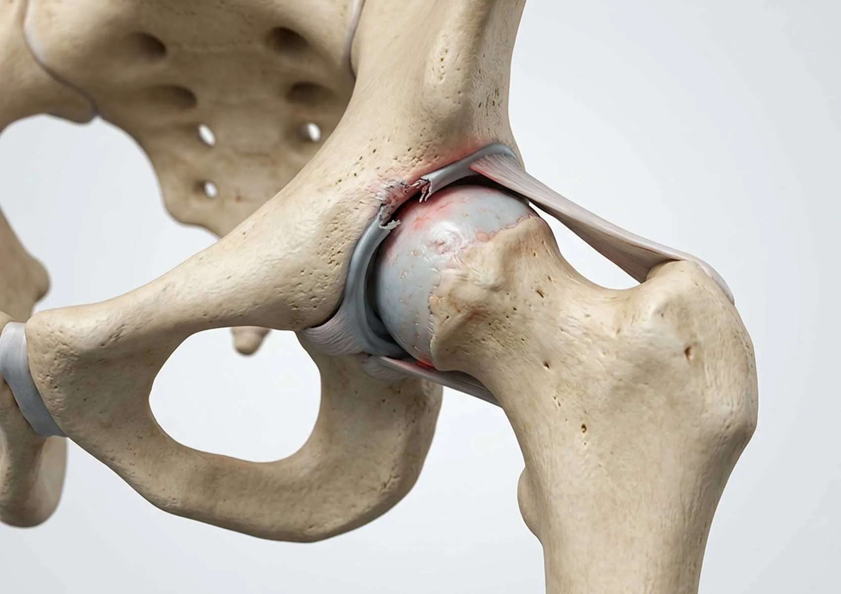
The hip joint is one of the most mobile and weight-bearing joints in the human body. However, sometimes a mismatch between the femoral head and the pelvic socket can cause abnormal friction during movement. Known in medical literature as femoroacetabular impingement (FAI), this condition is a common cause of hip pain especially among young and active individuals. If left undiagnosed early, this mechanical issue can lead to permanent damage to the joint cartilage and the labrum, the ring-like cartilage surrounding the socket.
Femoroacetabular impingement is not only a source of pain but also a significant risk factor for developing hip osteoarthritis later in life. Therefore, correct interpretation of symptoms and planning appropriate treatment are critical for preserving joint health. This article will explore the causes, symptoms, and current treatment approaches in detail.
What is Femoroacetabular Impingement?
Femoroacetabular impingement is a clinical condition caused by an anatomical mismatch between the femur (thigh bone) head and the acetabulum (hip socket). In a normal hip joint, the femoral head moves smoothly within the socket without friction. However, in FAI, bone deformities cause the bones to collide or pinch each other during movement. This can gradually damage the soft tissues inside the joint, such as the labrum and cartilage.
For more comprehensive information on the definition and treatment process of the condition, visit Definition and Treatment of Femoroacetabular Impingement. This mechanical problem can result from developmental differences in bone growth plates or be related to intense sports activity.
FAI occupies a special place among hip joint disorders and treatments because early intervention can preserve the joint’s natural structure. Untreated cases can progress to severe joint surface damage requiring more complex surgery.
Anatomical Causes and Types of Femoroacetabular Impingement
This syndrome is classified into different types based on the location of the bony deformity. Understanding anatomical variations is fundamental for determining treatment strategies. The diagram below visualizes these anatomical variations and impingement types of the hip joint.
Cam Type Impingement
Cam type impingement arises from deformity in the transition area between the femoral head and neck. Normally spherical, this region may be flattened or have a bump. This deformity causes the femoral head to jam against the acetabular rim when the hip is flexed, “grinding” the cartilage. It is more common in young males and athletes.
Pincer Type Impingement
Pincer type impingement involves excessive coverage of the femoral head by the acetabulum. This occurs when the socket is too deep or retroverted. During movement, the femoral neck impinges on the acetabular rim and labrum. This type is often seen in middle-aged women.
Mixed Type Impingement
The most common form in clinical practice, mixed type, features both Cam and Pincer deformities. Over 80% of cases exhibit both pathologies, with joint damage progressing faster due to their combined effects.
Symptoms of Hip Impingement and Its Impact on Quality of Life
Symptoms of femoroacetabular impingement usually start insidiously and intensify over time. Patients often describe the pain by making a “C” shape with their hand around the side of the hip. The pain is typically felt in the groin but can radiate to the outer thigh or buttocks.
The most characteristic symptom is sharp groin pain when standing up after prolonged sitting, getting out of a car, or squatting. Mechanical symptoms such as catching, locking, or a sensation of “pinching” during quick turns in sports may also occur. For detailed insights on how these symptoms affect daily life, see Hip Impingement Symptoms and Daily Life Impact. Pain can force patients to limit physical activity and may reduce overall quality of life.
Diagnostic Methods for Femoroacetabular Impingement
The diagnostic process begins with a thorough patient history and physical examination. The physician assesses the hip’s range of motion and applies specific tests to provoke impingement, such as the FADIR test. Pain during these maneuvers supports the suspicion of FAI.
Radiological imaging is essential for definitive diagnosis and bone structure evaluation. X-rays reveal Cam or Pincer deformities. To assess soft tissue damage, especially labral tears and cartilage status, Magnetic Resonance Imaging (MRI) or MR arthrography (contrast-enhanced MRI) may be used. Computed Tomography (CT) provides detailed three-dimensional views of bone anatomy and assists in surgical planning.
Treatment Options
Treatment of femoroacetabular impingement is individualized based on patient age, symptom severity, cartilage damage, and activity demands.
Conservative Treatment Methods
For patients with mild symptoms or minimal cartilage damage, non-surgical methods are tried first. These include activity modification to avoid painful movements, non-steroidal anti-inflammatory drugs (NSAIDs), and physical therapy. Physical therapy focuses on strengthening the muscles around the hip to balance joint loading and maintain mobility. In some cases, intra-articular injections can help control pain and are used diagnostically to confirm the hip as the pain source.
Surgical Options: Hip Arthroscopy
Surgery is considered for patients who do not respond to conservative treatment and experience primarily mechanical symptoms. Today, hip arthroscopy is the gold standard surgical method. Hip arthroscopy treatment options involve minimally invasive surgery using a camera and specialized instruments through small incisions.
During surgery, bony deformities causing the impingement are shaved down, and any labral tears are repaired. Arthroscopic treatment of femoroacetabular impingement restores the hip’s natural anatomy and eliminates impingement.
Postoperative Rehabilitation and Exercises
The success of surgery depends as much on postoperative rehabilitation as on surgical technique. Recovery varies by individual but typically includes the use of crutches and movement restrictions in the initial weeks. Gradual implementation of recommended exercises shown in the following visual series is essential.
Rehabilitation aims to restore joint range of motion and increase muscle strength. Rehabilitation processes in sports injuries should be followed diligently. Return to sports usually takes 4 to 6 months depending on the procedure.
Return-to-sport criteria are especially important for professional athletes. This rehabilitation approach is similar to that following anterior cruciate ligament (ACL) surgery, focusing on restoring muscle memory and proprioception (balance sense).
Risk Factors and Prevention Strategies
Genetic factors as well as high-intensity sports during growth periods (such as soccer, basketball, and ice hockey) are thought to contribute to the development of femoroacetabular impingement. Awareness of these risk factors is important to protect athlete health. Being informed about joint-preserving surgeries and patient selection can help prevent future osteoarthritis.
Improper training programs can also increase risk. Learning about common exercise mistakes in athletes is crucial, as they may overload the hip joint. General knowledge of injury risk factors and prevention in athletes supports preventative care. Flexibility training and core strengthening reduce joint load and help control symptoms.
Femoroacetabular impingement is a manageable condition with accurate diagnosis and modern treatments. Early intervention is the key to maintaining an active lifestyle and long-term hip health.
This content is provided for informational purposes only and does not constitute professional medical advice, diagnosis, or treatment. Always consult your physician or a qualified healthcare professional with any questions you may have regarding your health. Do not disregard medical advice or delay seeking care based on information provided on this site.
FAQ
-
What is femoroacetabular impingement (FAI)?
Femoroacetabular impingement is a condition where abnormal contact occurs between the head of the femur and the socket of the pelvis in the hip joint. This mechanical mismatch can cause pain, limited movement, and over time, damage to the cartilage.
-
What are the main symptoms of hip impingement?
The primary symptoms include groin or hip pain that worsens after prolonged sitting, driving, or squatting. There may also be restricted hip movement, a feeling of catching, and sometimes limping.
-
How is FAI diagnosed?
Diagnosis begins with a physical examination by a specialist and a review of the patient's symptoms. Confirmatory diagnosis involves imaging techniques such as X-rays, MRI, or CT scans to assess bone structure and soft tissues.
-
Can femoroacetabular impingement be treated?
Yes, it can be treated. Treatment options vary based on the patient's condition and include conservative methods like pain relievers, physical therapy, injections, or surgical interventions such as hip arthroscopy.
-
When should surgical treatment be considered?
Surgery is usually considered when conservative treatments (physical therapy, medication) fail to relieve persistent pain and when quality of life is significantly affected. The surgery aims to correct bone abnormalities causing impingement and repair any labral tears if present.