
Hip Arthroscopy: Comprehensive Guide and FAQs
Hip arthroscopy is a minimally invasive (closed) surgical technique used for diagnosing and treating hip joint problems.
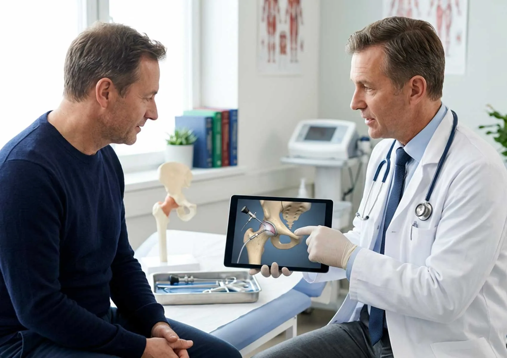
Hip arthroscopy is a minimally invasive treatment method increasingly used in orthopedics, enabled by advances in technology and surgical instruments. Commonly referred to as "closed hip surgery," this procedure allows visualization and treatment of pathology within the hip joint through small incisions of just a few millimeters, using a camera and specialized instruments. It plays a significant role in managing hip pain, especially among athletes and physically active individuals.
This surgical technique requires advanced expertise due to the deep anatomical position of the hip joint and the surrounding dense muscle layers, making it more technically demanding than knee or shoulder arthroscopy. Through hip arthroscopy, detailed examination of the joint cartilage, labrum (cartilage rim), ligaments, and joint capsule is possible. Compared to traditional open surgery, it offers potential benefits such as reduced pain, shorter hospital stays, and faster return to daily activities, contributing to its growing preference.
For more foundational information, see our article: What is Hip Arthroscopy?.
Introduction to Hip Arthroscopy
Historically, hip joint problems were managed primarily through open surgeries or conservative (non-surgical) treatments. However, with the advancement of imaging techniques such as MRI and CT scans and improvements in surgical methods, it has become clear that mechanical issues within the hip joint can often be corrected before degenerative changes (osteoarthritis) develop. Hip arthroscopy represents a cornerstone of these joint-preserving surgical approaches.
This procedure serves not only as a diagnostic tool but also as a therapeutic surgical intervention. Particularly for young and middle-aged adults where mechanical issues—such as bony overgrowths or tissue tears—are the cause of hip pain, arthroscopic treatment can address these lesions. The objective is to reduce pain, improve function, and preserve the natural joint for as long as possible.
Hip Joint Anatomy and Function
The hip is a ball-and-socket joint and one of the largest weight-bearing joints in the body. The femoral head (ball) fits into the acetabulum (socket) of the pelvis. Proper interaction between these structures allows pain-free movement with a wide range of motion.
A key structure for joint stability is the labrum, a ring of cartilage surrounding the acetabular rim. The labrum deepens the socket and acts as a seal, creating negative pressure (vacuum effect) inside the joint. This helps retain joint fluid, nourishing the cartilage and enhancing joint stability. The labrum is among the most frequently treated tissues during hip arthroscopy.
For details on healthy hip anatomy, please review our article on Hip Joint Anatomy.
What is Hip Arthroscopy?
Hip arthroscopy is a sophisticated endoscopic surgical procedure used to diagnose and treat hip joint disorders. It involves creating 2 to 4 small skin incisions of about 1 cm each (called portals). Through one portal, a thin tube called an arthroscope, equipped with a light source and camera, is inserted into the joint. The images captured are displayed in high resolution on a monitor. The surgeon views these images and operates using specialized instruments inserted through the other portals.
For technical details and step-by-step procedural information, visit our page on Hip Arthroscopy Procedure. For specific techniques used to treat impingement syndromes, our content on Arthroscopic Treatment of FAI is recommended.
How is it Performed?
Hip arthroscopy is typically performed under general or epidural anesthesia. Due to the hip joint’s tight and deep nature, controlled traction is applied to the leg using a specialized operating table to increase the joint space and allow the surgical instruments to maneuver freely. This process is continuously monitored via fluoroscopy (real-time X-ray).
The surgeon inspects the joint for damaged tissues. Torn labrum tissue can be repaired or cleaned (debrided). Cartilage damage may be treated through microfracture or grafting techniques. Bony overgrowths causing impingement (cam or pincer lesions) are shaved down using specialized burrs.
Advantages and Limitations
The primary advantage of hip arthroscopy is its minimally invasive nature. Compared to open surgery, there is significantly less trauma to muscles and soft tissues, resulting in reduced postoperative pain, shorter hospital stays (usually one day), and faster initiation of rehabilitation. Smaller incisions also result in more acceptable scarring from a cosmetic perspective.
However, limitations exist. In patients with advanced hip osteoarthritis where joint space is severely narrowed, arthroscopy can be technically challenging and outcomes may be limited. Additionally, in cases of hip dysplasia (developmental deformity), arthroscopy alone might not suffice, and supplemental surgical procedures may be necessary.
When is Hip Arthroscopy Indicated?
Hip arthroscopy is not suitable for all causes of hip pain. It is generally reserved for mechanical problems unresponsive to conservative treatments (medications, physical therapy, rest).
The most common indication is Femoroacetabular Impingement (FAI) Syndrome, where bony overgrowths on the femoral head or acetabular rim cause pinching during movement, damaging cartilage and the labrum. Arthroscopy removes these bony protrusions to relieve impingement. For more information, see FAI Treatment.
Other frequent indications include:
- Labral Tears: Repair of tears caused by trauma or impingement.
- Cartilage Damage: Treatment of focal cartilage defects.
- Synovial Diseases: Inflammation or tumorous formations of the synovial membrane (e.g., PVNS, synovial chondromatosis).
- Loose Bodies: Removal of cartilage or bone fragments floating inside the joint.
- Snapping Hip Syndrome: Release of tendons snapping over bony prominences.
For a comprehensive list of conditions, please review our pages on Hip Joint Disorders and Indications for Hip Arthroscopy.
Preoperative Evaluation and Preparation
A detailed preoperative assessment is crucial for successful surgery. Physical examination helps identify the source of pain. Radiological imaging (X-rays, MRI, and sometimes CT) provides detailed views of bone and soft tissue structures including the labrum and cartilage. In certain cases, intra-articular injection tests may be performed to confirm if pain originates from within the joint.
Once surgery is scheduled, anesthesia planning and preoperative precautions are arranged. Be sure to inform your physician about any blood-thinning medications, chronic illnesses, or allergies.
Postoperative Process and Recovery
Recovery time depends on the specific procedure performed (whether only cleaning or repair was done). Patients are often mobilized within hours after surgery. Where labral repair or cartilage procedures are involved, weight-bearing may be restricted for 2 to 4 weeks with crutches.
Physical Therapy and Exercises
Rehabilitation is vital to surgical success. The physical therapy program aims to maintain joint range of motion, restore muscle strength, and normalize gait patterns. Passive movements and isometric exercises dominate the early weeks, gradually advancing to strengthening and balance exercises.
Return to Daily Activities
Patients with desk jobs usually return to work within 2 to 3 weeks. Those engaged in physically demanding jobs may require 6 to 8 weeks. Driving is generally resumed when leg control is adequate and pain medication is discontinued, typically within 2 to 4 weeks. Return to sports varies between 4 to 6 months depending on the sport and surgical details. For professional athletes, this timeline is carefully managed with the guidance of a sports surgeon and physiotherapist.
Risks and Complications of Hip Arthroscopy
Like any surgery, hip arthroscopy carries potential risks, but complication rates are low in experienced hands. The most common temporary complication is numbness in the groin or thigh area due to traction-related nerve irritation (pudendal or lateral femoral cutaneous nerve neuropraxia), which usually resolves within weeks.
Rare complications include infection, bleeding, instrument breakage inside the joint, vascular or nerve injury, and deep vein thrombosis (blood clots). Preventive measures such as compression stockings and blood thinners may be prescribed postoperatively.
For more detailed information on possible complications, visit our page on Hip Arthroscopy Risks.
Comparing Hip Arthroscopy with Other Surgical Options
Hip arthroscopy is a joint-preserving surgery focused on maintaining the patient’s natural joint. For advanced osteoarthritis, total hip replacement surgery is performed, replacing the damaged joint with a prosthesis. Arthroscopy aims to delay or prevent the need for joint replacement by treating problems early.
Open hip surgery, such as for complex hip dislocation or deformity correction, provides full joint visualization but has longer recovery times and greater surgical trauma compared to arthroscopy. Open procedures are now reserved for highly complex cases where arthroscopy is inadequate.
If your hip damage is too advanced to be corrected arthroscopically, total hip replacement might be considered. Expectations and outcomes for this surgery can be reviewed in our article Life After Total Hip Replacement.
This content is provided for informational purposes only and does not constitute medical advice. It is essential to consult a qualified healthcare professional for diagnosis and treatment. The information presented here is not a substitute for professional medical evaluation. Please consult your physician if you have any questions regarding your medical condition. Do not delay seeking medical care based on the information provided.
FAQ
-
What is hip arthroscopy?
Hip arthroscopy is a minimally invasive surgical procedure that involves inserting a small camera and surgical instruments through tiny incisions to diagnose and treat problems within the hip joint.
-
Who is a suitable candidate for hip arthroscopy?
Patients with hip impingement, cartilage damage, torn labrum, or other intra-articular issues who have not responded to non-surgical treatments are typically suitable candidates. It's important to consult an orthopedic specialist for a precise evaluation.
-
How long does hip arthroscopy surgery take and what is the recovery like?
The surgery usually lasts between 1 to 2 hours depending on the complexity of the case. Recovery times vary per individual, but with physical therapy and exercises, most can return to daily activities within a few weeks.
-
What are the risks of hip arthroscopy surgery?
As with any surgery, there are risks including infection, bleeding, nerve injury, and blood clots. Your specialist will discuss these risks with you in detail.
-
What should be considered after hip arthroscopy?
It is crucial to closely follow your doctor's and physiotherapist’s instructions after surgery, perform regular exercises, and adhere to any restrictions. Pain management and wound care are also important during recovery.